

Read enough about metabolic research compounds and one acronym keeps surfacing: AMPK. It is offered as the explanation for why a peptide affects fat metabolism, why a small molecule is called an “exercise mimetic”, and why a drug prescribed to millions might also slow ageing. AMPK is a real and well-characterised enzyme, and most of that attention is deserved — but the shorthand hides a lot, and some of the claims attached to it do not survive contact with the primary literature.
This guide explains what AMPK is, how it is switched on, what it does when it is, and which of the compounds routinely called “AMPK activators” genuinely are. It spends as much space on the contested parts as on the settled ones.
The short answer
- AMPK is an enzyme, not a peptide. AMP-activated protein kinase is a protein kinase built from three different subunits, and it functions as the cell’s fuel gauge.
- It reads the adenine-nucleotide ratio. When a cell burns through ATP, AMP and ADP accumulate; AMP binding to AMPK’s gamma subunit activates the enzyme, while ATP keeps it switched off.
- Two upstream kinases arm the switch by phosphorylating a threonine in the alpha subunit, conventionally called Thr172: the tumour suppressor LKB1, and CaMKK2, which provides a calcium-triggered route.
- Once on, it flips the cell’s metabolic priorities: catabolic, ATP-generating processes up; anabolic, ATP-consuming ones down. Fatty-acid synthesis is inhibited through acetyl-CoA carboxylase; growth signalling is restrained through mTORC1; autophagy is switched on through ULK1.
- The metformin story is not settled. A 2001 paper reported that metformin activates AMPK; a 2010 paper found metformin still suppressed hepatic glucose production in cells with no AMPK at all. Reviews still describe the mechanism as debated.
- No direct AMPK activator is an approved medicine. The furthest any appears to have reached in people is an early-phase trial, and one pan-activator caused cardiac hypertrophy in animals.
- Not everything called an AMPK activator is one. MOTS-c research does implicate AMPK; SLU-PP-332 is an ERR agonist and 5-Amino-1MQ an NNMT inhibitor, and neither acts on AMPK.
AMP-activated protein kinase
AMPK · the cellular energy sensor · EC 2.7.11.1
- Molecule type
- Serine/threonine protein kinase — an enzyme, not a peptide or a receptor
- Architecture
- Heterotrimer: catalytic alpha (PRKAA1/PRKAA2), regulatory beta (PRKAB1/PRKAB2), regulatory gamma (PRKAG1/2/3)
- What it senses
- Adenine nucleotides at the gamma subunit: AMP activates, ATP inactivates; ADP also regulates
- Activating site
- Thr172 in the alpha subunit activation loop (numbered Thr183 in the alpha-1 isoform)
- Upstream kinases
- LKB1 (STK11) with STRADα and MO25; CaMKK2 on the calcium route
- Named by
- D. Grahame Hardie’s group, University of Dundee, 1989
- Approved activators
- None direct. Metformin is approved and activates AMPK indirectly, by a contested route
Where the evidence stands
- Cell studies
- Animal studies
- Early human trials of activators
- An approved AMPK-targeted medicine
AMPK’s biology is textbook material. Drugging it deliberately is not: the class remains investigational.
What AMPK is
AMPK is a protein kinase: an enzyme whose job is to attach phosphate groups to other proteins and so change what they do. Its full name, AMP-activated protein kinase, states its defining property. It is not a peptide, not a hormone and not a receptor, which matters because it is frequently discussed in the same breath as research peptides that are all three of those things.
Structurally it is a heterotrimer — three different subunits working as one unit. The catalytic alpha subunit does the phosphorylating and is encoded by either PRKAA1 or PRKAA2. A beta subunit (PRKAB1 or PRKAB2) scaffolds the complex. The gamma subunit (PRKAG1, PRKAG2 or PRKAG3) is the sensor: it binds adenine nucleotides, and which nucleotide is bound decides whether the enzyme is active. Because each slot has two or three options, mammals assemble twelve possible AMPK complexes, which is part of why activating “AMPK” is less simple than it sounds.
The sensing logic is elegant. ATP is the cell’s energy currency; spend it and you are left with ADP and AMP. AMP binding to the gamma subunit allosterically activates AMPK, while ATP promotes the inactive state, and structural work published in 2011 established that ADP also regulates the enzyme. So AMPK does not measure how much energy a cell has in absolute terms — it measures the ratio, which is a far more sensitive readout. Our guide to peptide receptors and GPCRs covers how cells read signals arriving from outside; AMPK is the intracellular counterpart, responding to metabolites instead.
How it was found
AMPK was not discovered as one thing. During the late 1970s, separate groups described kinase activities that inactivated two enzymes of lipid synthesis: HMG-CoA reductase and acetyl-CoA carboxylase. Both activities were stimulated by AMP, and for a decade it was unclear whether these were one enzyme or two.
The question was settled in 1989, when Carling, Clarke, Zammit and Hardie purified the enzyme from rat liver roughly 4,800-fold and showed that both kinase activities co-purified as a single AMP-activated enzyme, with a 63 kDa polypeptide carrying both the catalytic site and the AMP-binding capacity. AMP raised maximal activity three- to five-fold. That paper’s title uses the name the field has used ever since.
The upstream kinases took longer. In 2003, Woods and colleagues identified LKB1 — already known as a tumour suppressor mutated in Peutz-Jeghers syndrome — as the kinase that phosphorylates AMPK’s activation loop. Two years later, Hawley and colleagues showed that calcium could activate AMPK through a second route: in LKB1-deficient cells, calcium ionophores still raised AMPK phosphorylation, and depolarising rat brain slices activated AMPK without any measurable change in adenine nucleotide levels. That second route runs through CaMKK2, and it means AMPK is not purely an energy sensor; it is also a calcium sensor.
How the switch is thrown
Two inputs converge on the same enzyme, and in practice both are usually needed. The allosteric input is AMP binding to the gamma subunit, which directly raises activity and also protects the activating phosphate from being removed. The covalent input is phosphorylation of a threonine in the alpha subunit’s activation loop. The field calls this site Thr172, which is its number in the alpha-2 isoform; in the alpha-1 isoform the equivalent residue is numbered Thr183, so anyone checking a sequence database will meet both numbers for what is functionally one site. Two kinases put the phosphate there: LKB1, working in a complex with the pseudokinase STRADα and the scaffold MO25; and CaMKK2, triggered by a rise in intracellular calcium.
What it turns on, and off
The umbrella description is that activated AMPK boosts catabolic, ATP-generating pathways and shuts down anabolic, ATP-consuming ones. Underneath that umbrella sit specific, well-mapped substrates.
| Target | What AMPK does to it | Consequence reported |
|---|---|---|
| Acetyl-CoA carboxylase (ACC1, ACC2) | Phosphorylates and inhibits it | Fatty-acid synthesis falls, fatty-acid oxidation rises. Mice engineered so these sites cannot be phosphorylated show increased lipogenesis, insulin resistance and fatty liver |
| Raptor (within mTORC1) | Phosphorylates two conserved serines | mTORC1 is restrained; required for the cell-cycle arrest seen under energy stress |
| TSC2 | Phosphorylates and enhances its activity | A second brake on mTOR; required for translational and cell-size control |
| ULK1 and ULK2 | Phosphorylates them | Autophagy and mitophagy proceed. Losing AMPK or ULK1 causes defective mitophagy and p62 accumulation |
| PGC-1α | Phosphorylates it directly, at Thr177 and Ser538 | Mitochondrial and GLUT4 gene expression; in PGC-1α-deficient mice, AMPK’s effects on these genes largely disappeared |
| TBC1D1 | Phosphorylates it | Implicated in GLUT4 trafficking. In mouse muscle, contraction raised TBC1D1 phosphorylation in an AMPK-dependent way — though that study did not itself measure glucose uptake |
Skeletal muscle is the classic setting for all of this, because its energy demand swings more sharply between rest and work than almost any other tissue; a large review literature covers AMPK during exercise and recovery, in training adaptation and in substrate handling.
Activating AMPK in the laboratory
How you switch AMPK on in an experiment shapes what the experiment can conclude, and the available tools have real limitations.
| Tool | How it works | The caveat |
|---|---|---|
| AICAR | Converted inside the cell to an AMP mimetic; the long-standing method for activating AMPK in intact cells | Specificity was questioned in the very paper that established it — the 1995 title asks whether it is “a specific method” |
| A-769662 | The first widely used direct activator. Inhibited fatty-acid synthesis in rat hepatocytes; lowered plasma glucose by about 40% in ob/ob mice | Complex-selective, so it does not activate all twelve AMPK complexes |
| Compound 991 | A direct benzimidazole activator used in muscle research | Weak alone at 5 µM; its striking effect came only in combination with AICAR or with muscle contraction |
| PF-739 and PF-249 | PF-739 is a pan-activator; PF-249 is β1-selective. PF-739 lowered glucose in diabetic mice and in cynomolgus monkeys | The effect tracked muscle AMPK, not liver AMPK, and came from glucose disposal rather than reduced hepatic output. PF-249 activated hepatocyte AMPK and did not lower glucose at all |
| MK-8722 | A pan-activator reported to act on all twelve complexes; raised muscle glucose uptake independently of insulin in rodents and rhesus monkeys | Caused cardiac hypertrophy and cardiac glycogen accumulation — in the paper’s own title, and in primates as well as rodents |
| Genetic tools | AMPK-null and LKB1-null cells and mice; Thr172 and substrate-site knock-ins | The cleanest way to ask whether an effect needs AMPK, and the reason the metformin story turned out the way it did |
Most direct activators converge on the same allosteric pocket between the kinase domain and the carbohydrate-binding module, now generally called the ADaM site; a 2013 crystal structure of human AMPK showed a small-molecule activator bound there, stabilising the interaction between those two regions.
MOTS-c, NAD+ and related metabolic reagents are stocked as laboratory materials, priced in CAD.
Browse anti-aging & cellularThe metformin problem
This is the most instructive argument in the field, and it is a good illustration of the difference between a plausible mechanism and a demonstrated one.
In 2001, Zhou and colleagues reported in the Journal of Clinical Investigation that metformin activates AMPK in hepatocytes, with reduced acetyl-CoA carboxylase activity downstream, and proposed that AMPK activation gave “a unified explanation for the pleiotropic beneficial effects of this drug”. For most of the following decade that was the standard account, and it is still the version most often repeated outside the primary literature.
In 2010, Foretz and colleagues tested it the hard way, in the same journal, using hepatocytes lacking AMPK and hepatocytes lacking LKB1. If AMPK were the mechanism, metformin should have stopped working. Instead, cells lacking AMPK “displayed normal glucose production and gluconeogenic gene expression”, and metformin’s inhibition of glucose production was “amplified” in both AMPK- and LKB1-deficient cells, tracking a dose-dependent fall in intracellular ATP. Their conclusion was that metformin inhibits hepatic gluconeogenesis “in an LKB1- and AMPK-independent manner”.
Sixteen years later this is still not fully resolved, and current syntheses split the question rather than answering it. A 2021 review in Endocrine Reviews describes the mechanism by which metformin inhibits hepatic glucose production as “still highly debated”, and notes that the complex-I-inhibition-then-AMPK-activation account has “only been observed in the context of supra-pharmacological (>1 mM) metformin concentrations”. A 2026 review puts it more sharply: for metformin’s acute suppression of hepatic glucose production, “AMPK is therefore not considered fundamentally required”, while AMPK does appear to contribute to the drug’s chronic metabolic actions, mainly through ACC1 and ACC2 phosphorylation.
Why this matters when reading claims. “Compound X activates AMPK” and “compound X works because it activates AMPK” are different claims requiring different evidence. The first needs a phosphorylation readout. The second needs the effect to disappear when AMPK is removed. Most marketing copy, and a fair amount of review writing, treats the first as though it proved the second. Our guide to reading a peptide study covers this substitution in general terms.
Where peptides fit — and what isn’t AMPK
AMPK comes up constantly in research-compound writing, and it is worth separating the compounds whose published work genuinely implicates it from those where the association is loose or simply wrong.
MOTS-c: the AMPK link is real, and it is preclinical
MOTS-c is a 16-amino-acid peptide encoded inside the mitochondrial 12S rRNA gene — a peptide written in mitochondrial rather than nuclear DNA. In the 2015 Cell Metabolism paper that characterised it, Lee and colleagues reported that its cellular actions “inhibit the folate cycle and its tethered de novo purine biosynthesis, leading to AMPK activation”, with skeletal muscle as the primary target organ. In mice, MOTS-c treatment prevented age-dependent and high-fat-diet-induced insulin resistance, and diet-induced obesity.
So the AMPK connection is not invented. What it is not is clinical: that paper’s evidence is cell culture and mouse work, and it contains no human efficacy data. Our MOTS-c research guide covers the compound in detail, and MOTS-c, SS-31 and humanin places it alongside the other mitochondrial-derived peptides.
SLU-PP-332: an ERR agonist, not an AMPK activator
SLU-PP-332 is routinely grouped with AMPK activators under the heading of “exercise mimetics”. It is not one. It is a synthetic pan-agonist of the estrogen-related receptor family, acting on ERRα, ERRβ and ERRγ. ERRs are nuclear receptors — transcription factors that bind DNA and change which genes are read. AMPK is a kinase that phosphorylates proteins. These are different classes of target with different experimental signatures, and the published mouse work on SLU-PP-332 is about ERR agonism. Our SLU-PP-332 guide covers what that work actually showed.
5-Amino-1MQ: an NNMT inhibitor, upstream of NAD+
5-Amino-1MQ is also not an AMPK activator, and also not a peptide. It is a small methylquinolinium molecule that inhibits nicotinamide N-methyltransferase. NNMT methylates nicotinamide, diverting it away from NAD+ salvage; inhibit NNMT and intracellular NAD+ rises. That is a genuine metabolic intervention, but it runs through NAD+ availability rather than through the adenine-nucleotide sensing AMPK does. See our 5-Amino-1MQ guide and, for the coenzyme side, NAD+, NMN and nicotinamide riboside.
The cancer question, which cuts both ways
Because AMPK sits downstream of the tumour suppressor LKB1 and restrains anabolic growth, it was proposed to be a tumour suppressor. The evidence has not cooperated: AMPK also appears to help established tumours survive energy stress. Hardie’s 2015 review concluded that AMPK “can therefore be either a friend or a foe in cancer, depending on the context”, and a 2020 review put it as AMPK switching “from tumor suppression to tumor promotion” once tumorigenesis has occurred — implying activators might suit prevention while inhibitors might suit treating some cancers. Anyone presenting AMPK activation as unambiguously protective is overstating the record.
What’s established and what isn’t
Reasonably well established
- AMPK is a heterotrimeric kinase whose gamma subunit binds adenine nucleotides; AMP activates and ATP inactivates it.
- Activity requires phosphorylation of the alpha-subunit activation loop (Thr172) by LKB1 or CaMKK2.
- The calcium route through CaMKK2 can activate AMPK with no measurable change in nucleotide levels.
- AMPK inhibits acetyl-CoA carboxylase, restrains mTORC1 through raptor and TSC2, and switches on autophagy through ULK1.
- AMPK phosphorylates PGC-1α directly, at Thr177 and Ser538.
- No direct AMPK activator has been approved as a medicine; the class is investigational.
Open questions
- How much of metformin’s benefit depends on AMPK. Reviews still call the mechanism debated.
- Whether sustained pharmacological AMPK activation is safe. The pan-activator MK-8722 produced cardiac hypertrophy in rodents and primates.
- Which disease, if any, is the right target for AMPK activation. A 2019 review in Nature Reviews Drug Discovery states that the best indications and the long-term safety “remain to be established”.
- Whether AMPK restrains or assists tumours, which currently reads as context-dependent.
- Whether MOTS-c’s reported AMPK effects translate beyond cells and mice. No human efficacy data exist.
- Whether complex-selective activation of particular αβγ combinations avoids the problems pan-activation caused.
Sourcing metabolic reagents in Canada
If you are buying reagents for metabolic-signalling work, the documentation questions are the same as for any research peptide, with one addition specific to this area: several of the compounds involved are not peptides at all, and their certificates should reflect that.
- Check that identity testing suits the molecule. A 16-residue peptide such as MOTS-c is characterised by HPLC purity and a mass-spectrometry identity check; a small molecule like 5-Amino-1MQ, or a coenzyme like NAD+, is a different analytical problem. See our guides to reading an HPLC chromatogram and reading a mass spectrum.
- Match the batch number on the certificate to your vial. Our COA guide covers what else a useful certificate contains.
- Account for net peptide content. Synthetic peptides arrive as salts carrying counter-ions and water, so labelled mass is not peptide mass. Our guide to net peptide content works through it.
- Store cold, dry and dark, and keep material lyophilized until needed. See the peptide storage guide and what lyophilized means.
 MOTS-cThe mitochondrial-derived peptide whose 2015 paper reported AMPK activationView product
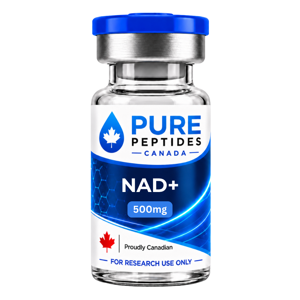 NAD+The redox coenzyme behind the adjacent, sirtuin-based energy-sensing pathwayView product
 5-Amino-1MQAn NNMT inhibitor — NAD+ salvage, not AMPKView product
 SLU-PP-332An ERR pan-agonist, frequently miscategorised as an AMPK activatorView product
Research use only. Pure Peptides Canada products are laboratory reagents for in-vitro research, not for human or veterinary use. This article explains published research for education; it is not medical, dietary or dosing guidance.
Frequently asked questions
What is AMPK?
AMPK, or AMP-activated protein kinase, is an enzyme that acts as the cell’s energy sensor. It is a protein kinase built from three subunits: a catalytic alpha subunit, a regulatory beta subunit and a regulatory gamma subunit that binds adenine nucleotides. AMP binding activates the enzyme and ATP keeps it inactive, so AMPK reads the ratio between them rather than any absolute energy level.
How is AMPK activated?
By two converging inputs. AMP binding to the gamma subunit allosterically activates the enzyme, and phosphorylation of a threonine in the alpha-subunit activation loop — conventionally called Thr172 — is required for full activity. Two upstream kinases add that phosphate: LKB1, working with STRAD-alpha and MO25, and CaMKK2, which responds to a rise in intracellular calcium and can activate AMPK without any measurable change in nucleotide levels.
What does AMPK do when it is switched on?
It reverses the cell’s metabolic priorities, turning on catabolic ATP-generating processes and turning off anabolic ATP-consuming ones. It phosphorylates and inhibits acetyl-CoA carboxylase, which lowers fatty-acid synthesis; it restrains mTORC1 through raptor and TSC2; it switches on autophagy and mitophagy through ULK1; and it phosphorylates PGC-1-alpha directly, at Thr177 and Ser538.
Is there an AMPK activator approved as a medicine?
No direct AMPK activator has been approved anywhere. Reviews describe the class as investigational, and the furthest any appears to have gone in people is an early-phase trial: the pan-activator O304 was tested in a proof-of-concept phase IIa study in type 2 diabetes. Metformin is an approved medicine that has been reported to activate AMPK, but it does so indirectly and by a route that is still debated.
Does metformin work by activating AMPK?
That was the standard account after a 2001 paper reported AMPK activation in hepatocytes, but it did not hold up as a complete explanation. In 2010, Foretz and colleagues found that metformin still suppressed glucose production in hepatocytes lacking AMPK or LKB1 — the effect was in fact amplified in those cells, and tracked a fall in intracellular ATP. Current reviews treat AMPK as contributing to some of metformin’s chronic effects while not being fundamentally required for the acute suppression of hepatic glucose production.
Is MOTS-c an AMPK activator?
The 2015 paper that characterised MOTS-c reported that its cellular actions inhibit the folate cycle and the de novo purine biosynthesis tethered to it, leading to AMPK activation, with skeletal muscle as the primary target organ. That evidence is cell-culture and mouse work; there is no human efficacy data in it. MOTS-c is also named on the 2026 World Anti-Doping Agency Prohibited List as an example of an AMPK activator.
Is SLU-PP-332 an AMPK activator?
No. SLU-PP-332 is a synthetic agonist of the estrogen-related receptor nuclear receptors, acting on ERR-alpha, ERR-beta and ERR-gamma. ERRs are transcription factors; AMPK is a protein kinase. The two are different classes of target and should not be conflated, even though both compounds are often filed under “exercise mimetics”. 5-Amino-1MQ is a third distinct case: an NNMT inhibitor that raises NAD+ availability and has no established action on AMPK.
Metabolic research reagents, documented by batch
MOTS-c, NAD+ and the rest of the metabolic range, priced in Canadian dollars and shipped tracked from inside Canada.
References
- Carling D, Clarke PR, Zammit VA, Hardie DG. Purification and characterization of the AMP-activated protein kinase: copurification of acetyl-CoA carboxylase kinase and 3-hydroxy-3-methylglutaryl-CoA reductase kinase activities. Eur J Biochem. 1989;186(1-2):129–136. DOI
- Corton JM, Gillespie JG, Hawley SA, Hardie DG. 5-Aminoimidazole-4-carboxamide ribonucleoside. A specific method for activating AMP-activated protein kinase in intact cells? Eur J Biochem. 1995;229(2):558–565. DOI
- Zhou G, Myers R, Li Y, et al. Role of AMP-activated protein kinase in mechanism of metformin action. J Clin Invest. 2001;108(8):1167–1174. DOI
- Inoki K, Zhu T, Guan K-L. TSC2 mediates cellular energy response to control cell growth and survival. Cell. 2003;115(5):577–590. DOI
- Woods A, Johnstone SR, Dickerson K, et al. LKB1 is the upstream kinase in the AMP-activated protein kinase cascade. Curr Biol. 2003;13(22):2004–2008. DOI
- Hawley SA, Pan DA, Mustard KJ, et al. Calmodulin-dependent protein kinase kinase-β is an alternative upstream kinase for AMP-activated protein kinase. Cell Metab. 2005;2(1):9–19. DOI
- Cool B, Zinker B, Chiou W, et al. Identification and characterization of a small molecule AMPK activator that treats key components of type 2 diabetes and the metabolic syndrome. Cell Metab. 2006;3(6):403–416. DOI
- Jäger S, Handschin C, St-Pierre J, Spiegelman BM. AMP-activated protein kinase (AMPK) action in skeletal muscle via direct phosphorylation of PGC-1α. Proc Natl Acad Sci USA. 2007;104(29):12017–12022. DOI
- Gwinn DM, Shackelford DB, Egan DF, et al. AMPK phosphorylation of raptor mediates a metabolic checkpoint. Mol Cell. 2008;30(2):214–226. DOI
- Pehmøller C, Treebak JT, Birk JB, et al. Genetic disruption of AMPK signaling abolishes both contraction- and insulin-stimulated TBC1D1 phosphorylation and 14-3-3 binding in mouse skeletal muscle. Am J Physiol Endocrinol Metab. 2009;297(3):E665–E675. DOI
- Foretz M, Hébrard S, Leclerc J, et al. Metformin inhibits hepatic gluconeogenesis in mice independently of the LKB1/AMPK pathway via a decrease in hepatic energy state. J Clin Invest. 2010;120(7):2355–2369. DOI
- Xiao B, Sanders MJ, Underwood E, et al. Structure of mammalian AMPK and its regulation by ADP. Nature. 2011;472(7342):230–233. DOI
- Egan DF, Shackelford DB, Mihaylova MM, et al. Phosphorylation of ULK1 (hATG1) by AMP-activated protein kinase connects energy sensing to mitophagy. Science. 2011;331(6016):456–461. DOI
- Fullerton MD, Galic S, Marcinko K, et al. Single phosphorylation sites in Acc1 and Acc2 regulate lipid homeostasis and the insulin-sensitizing effects of metformin. Nat Med. 2013;19:1649–1654. DOI
- Xiao B, Sanders MJ, Carmena D, et al. Structural basis of AMPK regulation by small molecule activators. Nat Commun. 2013;4:3017. DOI
- Lee C, Zeng J, Drew BG, et al. The mitochondrial-derived peptide MOTS-c promotes metabolic homeostasis and reduces obesity and insulin resistance. Cell Metab. 2015;21(3):443–454. DOI
- Hardie DG. Molecular pathways: is AMPK a friend or a foe in cancer? Clin Cancer Res. 2015;21(17):3836–3840. DOI
- Bultot L, Lai Y-C, Jensen TE, et al. Benzimidazole derivative small-molecule 991 enhances AMPK activity and glucose uptake induced by AICAR or contraction in skeletal muscle. Am J Physiol Endocrinol Metab. 2016;311(4):E706–E719. DOI
- Cokorinos EC, Delmore J, Reyes AR, et al. Activation of skeletal muscle AMPK promotes glucose disposal and glucose lowering in non-human primates and mice. Cell Metab. 2017;25(5):1147–1159.e10. DOI
- Myers RW, Guan H-P, Ehrhart J, et al. Systemic pan-AMPK activator MK-8722 improves glucose homeostasis but induces cardiac hypertrophy. Science. 2017;357(6350):507–511. DOI
- Neelakantan H, Vance V, Wetzel MD, et al. Selective and membrane-permeable small molecule inhibitors of nicotinamide N-methyltransferase reverse high fat diet-induced obesity in mice. Biochem Pharmacol. 2018;147:141–152. DOI
- Kjøbsted R, Hingst JR, Fentz J, et al. AMPK in skeletal muscle function and metabolism. FASEB J. 2018;32(4):1741–1777. DOI
- Steneberg P, Lindahl E, Dahl U, et al. PAN-AMPK activator O304 improves glucose homeostasis and microvascular perfusion in mice and type 2 diabetes patients. JCI Insight. 2018;3(12):e99114. DOI
- Steinberg GR, Carling D. AMP-activated protein kinase: the current landscape for drug development. Nat Rev Drug Discov. 2019;18(7):527–551. DOI
- Vara-Ciruelos D, Dandapani M, Hardie DG. AMP-activated protein kinase: friend or foe in cancer? Annu Rev Cancer Biol. 2020;4(1):1–16. DOI
- LaMoia TE, Shulman GI. Cellular and molecular mechanisms of metformin action. Endocr Rev. 2021;42(1):77–96. DOI
- Billon C, Schoepke E, Avdagic A, et al. A synthetic ERR agonist alleviates metabolic syndrome. J Pharmacol Exp Ther. 2024;388(2):232–240. DOI
- Ceballos H, Zhang E, Huang W. AMPK therapy: a little goes a long way. Cells. 2026;15(16):1503. DOI
- UniProt Consortium. UniProtKB entries P54646 (PRKAA2) and Q13131 (PRKAA1), human AMPK catalytic subunits alpha-2 and alpha-1.
- IUBMB Enzyme Nomenclature. EC 2.7.11.31, [hydroxymethylglutaryl-CoA reductase (NADPH)] kinase, with its 1978 historical references.
For laboratory research use only. Not for human or veterinary use. This article summarizes published research for educational purposes and is not medical advice.