

One human gene produces a protein that is then cut into around a dozen separate signalling molecules — a stress hormone, an opioid, and a peptide best known for making skin darker. That last one is α-MSH, and it is a more interesting molecule than its nickname suggests. The same thirteen residues that drive pigment synthesis in a melanocyte also act as one of the body’s own anti-inflammatory signals, and the last three of those residues are sold in laboratories as a research peptide in their own right.
This guide explains where α-MSH comes from, what the five melanocortin receptors actually respond to (they are not interchangeable, and one of them ignores α-MSH entirely), how the pigment switch works, what the anti-inflammatory evidence does and does not show, and where the KPV fragment sits in all of it.
The short answer
- α-MSH is a 13-residue peptide: Ser-Tyr-Ser-Met-Glu-His-Phe-Arg-Trp-Gly-Lys-Pro-Val, acetylated at the N-terminus and amidated at the C-terminus.
- It is literally the first 13 residues of ACTH. Both are cut from proopiomelanocortin (POMC) starting at the same residue, so α-MSH is ACTH(1–13) with modified ends.
- POMC processing is tissue-dependent. One convertase, PC1, yields ACTH in the anterior pituitary; a second, PC2, cleaves ACTH further to α-MSH in the intermediate lobe. “POMC becomes α-MSH” skips a step and a tissue.
- There are five melanocortin receptors, all Gs-coupled — but MC2R responds only to ACTH, not to any MSH. And α-MSH is ranked last among endogenous agonists at MC3R.
- MC1R loss of function does not mean no pigment. It means a different pigment: the switch runs from eumelanin toward the red-yellow, pro-oxidant pheomelanin.
- α-MSH also inhibits NF-κB in cultured cells, selectively — one 1998 study found it blocked TNF-induced NF-κB activation while leaving AP-1 untouched.
- KPV is α-MSH residues 11–13 — Lys-Pro-Val. Whether it works through a melanocortin receptor is unsettled, and the published work on it is cells and mice.
- Two melanocortin medicines are authorized abroad: setmelanotide, an MC4R agonist, and afamelanotide, an α-MSH analogue for a rare photodermatosis. Neither is a research peptide, and both are used under medical supervision.
α-Melanocyte-stimulating hormone
α-MSH · alpha-melanotropin · ACTH(1–13) amide
- Length
- 13 amino acids
- Sequence
- Ac-Ser-Tyr-Ser-Met-Glu-His-Phe-Arg-Trp-Gly-Lys-Pro-Val-NH2
- Precursor
- Proopiomelanocortin (POMC), a 267-residue protein; α-MSH is POMC residues 138–150
- Terminal modifications
- N-acetylserine at residue 1; valine amide at residue 13
- Sibling peptides from POMC
- ACTH, β-MSH, γ-MSH, CLIP, β-lipotropin, β-endorphin, Met-enkephalin
- Receptors
- MC1R, MC3R, MC4R, MC5R — all Gs-coupled. Not MC2R, which recognises only ACTH
- Preferred at
- MC1R, where α-MSH is the top-ranked endogenous agonist
- Endogenous antagonists
- Agouti signalling protein (ASIP) and agouti-related peptide (AGRP)
- Known fragment in research use
- KPV — residues 11–13
Where the evidence stands
- Cell studies
- Animal studies
- Approved melanocortin medicines (MC4R and MC1R agonists)
- Human clinical data for the KPV fragment
The receptor pharmacology is solid and two analogues are authorized medicines. The research-peptide fragment is a different and much thinner story.
What α-MSH is, and where it comes from
α-MSH is a thirteen-residue peptide, and the cleanest way to understand it is to look at what it is cut from. Proopiomelanocortin — POMC — is a 267-residue precursor protein that functions as a kind of hormone warehouse. Depending on which enzymes are present in which tissue, it yields adrenocorticotropic hormone, the three melanocyte-stimulating hormones, corticotropin-like intermediary peptide, β-lipotropin, γ-lipotropin, β-endorphin and Met-enkephalin. One gene; a stress hormone and an endogenous opioid and a pigment signal.
The residue coordinates make one relationship unmissable. In the curated human POMC record, ACTH spans residues 138 to 176, and α-MSH spans 138 to 150. They start at the same place. α-MSH is the N-terminal thirteen residues of ACTH, with an acetyl group added to the first serine and the C-terminal valine converted to an amide. Those two modifications are not cosmetic: they are part of what makes α-MSH a distinct signalling molecule rather than a degradation product.
The processing is also tissue-dependent, which is where most short summaries go wrong. Prohormone convertase 1 (PCSK1) acting in the anterior pituitary generates ACTH and β-lipotropin. It takes a second enzyme, prohormone convertase 2, in the intermediate lobe, to cleave ACTH again into α-MSH plus CLIP. So “POMC is cleaved into α-MSH” compresses two enzymes and two anatomical compartments into one arrow.
POMC is expressed well beyond the pituitary — in the hypothalamus, skin, lung, gut and pancreas — which is often cited to argue for locally made α-MSH acting as a paracrine signal in skin. One caution from the same literature is worth carrying: non-pituitary POMC transcripts are frequently not full length, so peptide levels outside the pituitary are low and their function is not clear. For a broader view of how precursor processing shapes peptide signalling, see our guide to peptide hormones.
The five melanocortin receptors
Melanocortins act through five G-protein-coupled receptors, MC1R through MC5R, and the pharmacology database lists the Gs family as the principal transduction mechanism for all five. That means the canonical route applies throughout: receptor to Gs, Gs to adenylyl cyclase, cAMP to protein kinase A. Our guides to GPCRs and peptide receptors and cyclic AMP and protein kinase A cover that machinery in detail.
What is not uniform is which ligand each receptor prefers — and this is where the most common error in melanocortin writing lives.
| Receptor | Endogenous agonist rank order | Where it is found |
|---|---|---|
| MC1R | α-MSH > β-MSH > ACTH > γ-MSH | Melanocytes, keratinocytes, dermal fibroblasts, immune cells including neutrophils, monocytes, macrophages and B lymphocytes, endothelial cells, dendritic cells, glia |
| MC2R | ACTH only — it does not recognise any MSH | Adrenal cortex, adipose tissue, bone cells |
| MC3R | γ-MSH > β-MSH > ACTH > α-MSH last | Central and peripheral |
| MC4R | β-MSH > α-MSH > ACTH > γ-MSH | Central and peripheral; brain MC4R is involved in hunger, satiety and energy expenditure |
| MC5R | α-MSH > β-MSH > ACTH > γ-MSH | Central and peripheral |
Two consequences follow. First, any sentence of the form “α-MSH acts on the melanocortin receptors MC1–MC5” is wrong at MC2R, which the pharmacology database describes as recognising only the ACTH peptide and which the protein record names outright as the adrenocorticotropic hormone receptor. Loss of MC2R function causes familial glucocorticoid deficiency. Second, α-MSH is ranked last among the four endogenous agonists at MC3R, so the fairly common claim that α-MSH resolves inflammation “via MC3R” overstates a weak ligand pairing.
The system also has endogenous brakes, which is unusual and worth knowing. Agouti signalling protein (ASIP) and agouti-related peptide (AGRP) oppose melanocortin signalling. One precision point: ASIP binds MC1 and MC4 with high affinity in vitro, but because it is normally expressed only in skin, physiologically it reaches only MC1 — a nice illustration of the difference between what a molecule can bind and what it ever meets. AGRP targets MC3 and MC4. The database is itself inconsistent about whether to call these antagonists or inverse agonists, describing them one way in its general text and another in its pharmacology section, so “endogenous antagonists” is the safe phrasing.
The pigment switch
The classic α-MSH story is pigmentation, and the mechanism is a textbook cAMP cascade. α-MSH binds MC1R on a melanocyte; adenylyl cyclase raises cAMP; PKA phosphorylates CREB; CREB activates the promoter of MITF, the master melanocyte transcription factor; MITF upregulates tyrosinase and the related enzymes TYRP1 and DCT; and eumelanin synthesis increases.
The part that gets garbled is what happens when MC1R does not work. Loss of MC1R function does not mean an absence of pigment — it means a different pigment. The switch defaults toward pheomelanin, the red-yellow pigment, which shields poorly against ultraviolet radiation and can amplify UVA-induced reactive oxygen species. That is the biological content behind the familiar association between red hair, fair skin and poor tanning.
That association traces to a 1995 letter in Nature Genetics, which reported MC1R variants in over 80% of individuals with red hair or fair skin that tans poorly, in fewer than 20% of individuals with brown or black hair, and in less than 4% of those with a good tanning response, concluding that MC1R is a control point in the regulation of pigmentation phenotype. The specific variant nomenclature came later: the field now distinguishes strongly associated “R” or RHC alleles (D84E, R142H, R151C, R160W, D294H) from more weakly associated “r” alleles (V60L, V92M, R163Q), and they differ mechanistically — some reduce cell-surface expression, others impair coupling. MC1R is highly polymorphic, with more than 200 variants described.
There is also direct human evidence that MC1R activation pigments skin without any ultraviolet exposure, and it comes from an unexpected place: the current prescribing information for the MC4R agonist setmelanotide notes that the MC1 receptor is expressed on melanocytes and that activating it leads to melanin accumulation and increased skin pigmentation independently of ultraviolet light — documented there as a drug side effect, because setmelanotide has residual MC1R activity.
One tidy inference that does not hold: a 2026 study tested nine melanocortin agonists against thirty MC1R variants and then in LPS-stimulated whole blood from genotyped volunteers, and found that loss of function presents as a continuum rather than a binary, that most compounds’ anti-inflammatory efficacy was diminished in variant carriers — and that Fitzpatrick skin type did not predict drug response. The authors explicitly advise against using red hair or skin type as a proxy for melanocortin pharmacology.
The other arm: inflammation
The reason α-MSH appears in research-peptide writing at all is not pigment. It is that MC1R is expressed on immune cells — neutrophils, monocytes, macrophages, B lymphocytes, dendritic cells — and that α-MSH behaves as an endogenous anti-inflammatory signal.
The best-characterised single result is a 1998 study in The Journal of Immunology, which reported that α-MSH blocked TNF-induced NF-κB activation in a dose- and time-dependent manner and also suppressed activation induced by LPS, okadaic acid and ceramide. It used four readouts — DNA binding by mobility shift assay, IκBα degradation, p65 nuclear translocation, and an NF-κB reporter — and the effect appeared to be cAMP-mediated, since adenylate cyclase and PKA inhibitors reversed it and a cAMP analogue mimicked it. Crucially, TNF-induced AP-1 activation was unaffected: this was selective, not a general shutdown. It is also cell-culture work, and it is frequently cited as though it had established α-MSH as a clinical anti-inflammatory, which it did not. Our NF-κB guide sets out what that pathway is and why a phospho-p65 result is narrower than it sounds.
Review literature adds a broader picture — suppression of TNF-α, IFN-γ, IL-1, IL-6 and IL-8 with induction of IL-10, reduced nitric oxide and prostaglandin output, regulatory T-cell induction, enhanced efferocytosis — but almost all of it is cells and mice. The human material in those reviews is observational: α-MSH measured in bronchoalveolar lavage fluid, or in synovial fluid, with associations rather than interventions.
And the receptor question is genuinely open. A 2023 review dedicated to melanocortins in inflammatory bowel disease found MC1R the best-supported candidate — mice with a mutant MC1R developed more severe colitis than wild-type — while noting that the MC3R evidence comes from non-intestinal mouse models, that MC3R and MC4R expression in human intestinal tissue is a correlation rather than a causal role, and that the roles of MC3R and MC4R in that disease remain mainly unexplored. The review does not cleanly assign α-MSH’s anti-inflammatory action to one receptor, and neither should anyone else.
KPV: the fragment
KPV is Lys-Pro-Val: α-MSH residues 11, 12 and 13, corresponding to POMC 148–150. It is about as small as a bioactive peptide gets, and its appeal in the literature is that it appears to carry anti-inflammatory activity without pigmentary effects.
A small accuracy note, because it propagates: a peer-reviewed 2023 review calls KPV “α-MSH(11–14)”, which cannot be right — three residues cannot span four positions. Checked against the curated POMC sequence, Lys-Pro-Val is residues 11 to 13 of the thirteen-residue hormone.
What the published work shows, stated at the level the sources support:
| Question | What the literature actually supports |
|---|---|
| Does KPV reduce inflammation in animals? | Yes, in mouse colitis models. A 2008 study in Inflammatory Bowel Diseases reported faster recovery, more weight regained, fewer histological infiltrates and lower colonic myeloperoxidase activity, and rescued all animals from death in a group with non-functional MC1R. Readouts were weight, histology and myeloperoxidase — not NF-κB |
| Does it act through a melanocortin receptor? | Unsettled, and two reviews read the same mouse data differently. The original authors concluded the effect was at least partly MC1R-independent; a 2023 review re-reads that experiment more conservatively and concludes KPV likely uses both MC1R-dependent and independent routes. Separate cell work in human bronchial epithelial cells implicates MC3R |
| How does it get into cells? | Proposed, not demonstrated. The 2008 Gastroenterology paper’s title asserts PepT1-mediated uptake; a 2023 review still describes this as hypothesised. The transporter (SLC15A1) does carry two-to-four-residue peptides, but with an annotated preference for dipeptides, and its annotated tissue is small intestine |
| Is there human clinical data? | None that we could locate. Two dedicated reviews of melanocortins in inflammatory bowel disease report no human clinical trial data for KPV or α-MSH in that setting, describing human experience with melanocortin agents as primarily anecdotal |
The inference worth resisting is the one the citation trail invites: evidence for the parent hormone is not evidence for the fragment. α-MSH has multi-assay NF-κB data; KPV has mouse efficacy data and a proposed transporter. Both are interesting. They are not the same claim. Our KPV research guide covers the compound in depth, and short peptides explained covers why a tripeptide behaves so unlike the molecule it came from.
KPV and the KPV-containing blends are stocked as laboratory reagents, priced in CAD.
Browse recovery & repairThe melanocortin medicines
Two authorized medicines act on this system, and they are useful calibration for anyone reading claims about melanocortin research compounds. Both are prescription products used under medical supervision; nothing here describes or recommends their use.
Setmelanotide is an MC4R agonist — its label describes roughly twentyfold less activity at MC3R and MC1R. It is approved to reduce excess body weight and maintain weight reduction in patients with acquired hypothalamic obesity, with Bardet-Biedl syndrome, or with obesity due to POMC, PCSK1 or LEPR deficiency confirmed by genetic testing. There is a point of real mechanistic interest here that is easy to get wrong: setmelanotide is an MC4R agonist but is not indicated for MC4R deficiency. Every approved genetic indication is a lesion upstream of an intact MC4R — and two of the three named genes, POMC and PCSK1, are precisely the precursor and the convertase from the first section of this article. An agonist cannot rescue a receptor that does not work.
Afamelanotide is a structural analogue of α-MSH that binds predominantly to MC1R. It differs from the native hormone at just two positions — norleucine for methionine at residue 4, and D-phenylalanine for L-phenylalanine at residue 7 — and those two substitutions raise potency by a reported ten- to thousandfold depending on the assay, with greater resistance to serum and proteolytic degradation. It was first authorized in the European Union on 22 December 2014 and by the US Food and Drug Administration on 8 October 2019, supplied as a subcutaneous implant for a rare light-sensitivity disorder, erythropoietic protoporphyria. The two regulators word the indication differently — the FDA’s is to increase pain-free light exposure in adults with a history of phototoxic reactions, the EMA’s is prevention of phototoxicity — so the two are not interchangeable quotations.
We will note what we could not verify rather than fill the gap: afamelanotide is widely equated with grey-market “Melanotan I”, and we found no primary or regulator source establishing that equivalence. The word “Melanotan” does not appear in the EMA’s assessment report at all. This article does not cover tanning peptides, and we do not provide research framing for pigmentation agonists; the regulatory position on unauthorized injectable products sold online is set out in the status note below.
What’s established and what isn’t
Reasonably well established
- α-MSH is POMC residues 138–150, identical to ACTH(1–13), acetylated at the N-terminus and amidated at the C-terminus.
- POMC processing is tissue-dependent: PC1 yields ACTH in the anterior pituitary, PC2 cleaves ACTH to α-MSH in the intermediate lobe.
- All five melanocortin receptors couple principally to Gs; MC2R recognises only ACTH.
- α-MSH is the top-ranked endogenous agonist at MC1R and the lowest-ranked at MC3R.
- MC1R activation drives eumelanin through cAMP, PKA, CREB, MITF and tyrosinase; loss of function defaults toward pheomelanin.
- MC1R variants are strongly associated with red hair and poor tanning, and the gene is highly polymorphic.
- α-MSH inhibits TNF-induced NF-κB activation in cultured cells, selectively and apparently via cAMP.
- KPV is α-MSH(11–13) and reduces inflammation in mouse colitis models.
- Two melanocortin receptor agonists are authorized medicines abroad.
Open questions
- Which receptor mediates α-MSH’s anti-inflammatory effects. MC1R has the most support in colitis models; MC3R and MC4R remain largely unexplored there.
- Whether KPV acts through a melanocortin receptor, independently of one, or both. Two reviews read the same mouse experiment differently.
- Whether PepT1 really transports KPV, or whether that remains a hypothesis.
- Whether KPV does anything measurable in humans. We could locate no published human clinical trial data.
- How much α-MSH is actually made outside the pituitary, given that peripheral POMC transcripts are often not full length.
- Whether grey-market “Melanotan I” is chemically the same molecule as afamelanotide. We found no regulator or primary source establishing it, and do not assume it.
- α-MSH’s circulating half-life as a number. Regulator documents describe afamelanotide as having a significantly increased half-life relative to the native hormone, but we found no figure for α-MSH itself.
Sourcing melanocortin reagents in Canada
If you are buying material for melanocortin-pathway work, the documentation questions are the standard ones, with two that matter particularly here.
- Identity testing matters more than usual for short peptides. A tripeptide has a small mass and few distinguishing features, so a mass-spectrometry identity check is doing real work. See reading a mass spectrum and reading an HPLC chromatogram.
- Watch endotoxin if you are measuring inflammatory readouts. LPS is itself a canonical activator of the pathways these experiments look at, so carried-over endotoxin can imitate a result. Our note on endotoxin and research peptides covers what to ask.
- Match the batch number on the certificate to your vial — our COA guide covers what else a useful certificate contains.
- Account for net peptide content, since synthetic peptides arrive as salts carrying counter-ions and water: net peptide content explained.
- Store cold, dry and dark, and keep material lyophilized until needed: the storage guide and what lyophilized means.
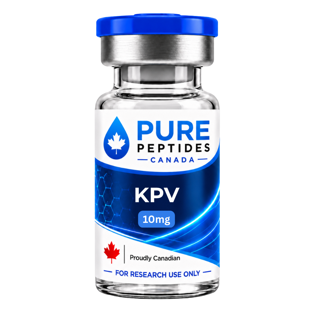 KPVα-MSH residues 11–13, the fragment in the colitis literatureView product
 GHK-Cu + KPV BlendThe KPV tripeptide co-formulated with the copper tripeptideView product
 KLOW BlendA multi-component research blend that includes KPVView product
Research use only. Pure Peptides Canada products are laboratory reagents for in-vitro research, not for human or veterinary use. This article explains published research for education; it is not medical, dietary or dosing guidance.
Frequently asked questions
What is alpha-MSH?
Alpha-melanocyte-stimulating hormone is a 13-amino-acid peptide cut from the precursor protein proopiomelanocortin, or POMC. Its sequence is Ser-Tyr-Ser-Met-Glu-His-Phe-Arg-Trp-Gly-Lys-Pro-Val, with the N-terminal serine acetylated and the C-terminal valine amidated. In the curated human POMC record it occupies residues 138 to 150, which makes it identical to the first 13 residues of ACTH, since ACTH begins at the same residue. It acts through melanocortin receptors, which are G-protein-coupled receptors that raise cyclic AMP.
What does alpha-MSH do?
It has two well-studied arms. The one it is named for is pigmentation: acting on MC1R on melanocytes, it raises cyclic AMP, which through PKA, CREB and the transcription factor MITF increases tyrosinase and drives synthesis of eumelanin. The second arm is immune. MC1R is expressed on neutrophils, monocytes, macrophages and B lymphocytes, and alpha-MSH behaves as an endogenous anti-inflammatory signal; a 1998 study reported that it blocked TNF-induced NF-kB activation in cultured cells while leaving AP-1 activation unaffected. Most of the anti-inflammatory evidence is cell and animal work.
What are the melanocortin receptors?
Five G-protein-coupled receptors, MC1R to MC5R, all of which couple principally to Gs and therefore raise cyclic AMP. They are not interchangeable. MC1R is the melanocyte and immune-cell receptor, and alpha-MSH is its top-ranked endogenous agonist. MC2R responds only to ACTH and to no MSH at all, which is why it is also called the ACTH receptor. MC3R’s preferred endogenous ligand is gamma-MSH, with alpha-MSH ranked last of the four. MC4R in the brain is involved in hunger, satiety and energy expenditure. The system also has endogenous antagonists: agouti signalling protein and agouti-related peptide.
Is KPV the same as alpha-MSH?
No. KPV is a three-residue fragment of it: lysine, proline and valine, which are residues 11 to 13 of the 13-residue hormone. It is often described as carrying anti-inflammatory activity without pigmentary effects. The important distinction when reading claims is that evidence for the parent hormone is not evidence for the fragment. Alpha-MSH has multi-assay NF-kB data in cultured cells; KPV has mouse colitis efficacy data and a proposed transport route via PepT1 that a 2023 review still describes as hypothesised. Note also that one peer-reviewed review miscalls KPV “alpha-MSH(11-14)”; three residues cannot span four positions, and the correct range is 11 to 13.
Why does MC1R affect hair and skin colour?
Because MC1R signalling decides which pigment a melanocyte makes. When alpha-MSH activates MC1R, cyclic AMP rises, PKA phosphorylates CREB, CREB activates the MITF promoter, and MITF upregulates tyrosinase and related enzymes to produce eumelanin. When MC1R function is reduced, the balance shifts toward pheomelanin, the red-yellow pigment, which shields poorly against ultraviolet radiation. So loss of MC1R function does not mean no pigment; it means a different and less protective pigment. A 1995 report found MC1R variants in over 80% of people with red hair or poorly tanning fair skin, compared with under 4% of those who tanned well.
Is there an approved alpha-MSH analogue?
Yes. Afamelanotide is a structural analogue of alpha-MSH that binds predominantly to MC1R, differing from the native hormone at only two positions: norleucine in place of methionine at residue 4, and D-phenylalanine in place of L-phenylalanine at residue 7. It was authorized in the European Union on 22 December 2014 and by the US Food and Drug Administration on 8 October 2019, as a subcutaneous implant for erythropoietic protoporphyria, a rare light-sensitivity disorder. A second melanocortin medicine, setmelanotide, is an MC4R agonist approved for specific genetic and acquired forms of obesity. Both are prescription products used under medical supervision.
Does alpha-MSH reduce inflammation?
In cultured cells and in animal models, there is substantial evidence that it does, and the mechanism studied most directly is inhibition of NF-kB activation. The 1998 study most often cited used four separate readouts and found the effect was selective, since TNF-induced AP-1 activation was not affected, and appeared to depend on cyclic AMP. Review literature adds suppression of several pro-inflammatory cytokines with induction of IL-10. What is not established is which receptor mediates the effect: MC1R has the most support in colitis models, while a 2023 review describes the roles of MC3R and MC4R there as mainly unexplored. The human evidence in those reviews is observational rather than interventional.
Melanocortin research reagents, documented by batch
KPV and the KPV-containing blends, priced in Canadian dollars and shipped tracked from inside Canada.
References
- Valverde P, Healy E, Jackson I, Rees JL, Thody AJ. Variants of the melanocyte-stimulating hormone receptor gene are associated with red hair and fair skin in humans. Nat Genet. 1995;11(3):328–330. DOI
- Lipton JM, Catania A. Anti-inflammatory actions of the neuroimmunomodulator α-MSH. Immunol Today. 1997;18(3):140–145. DOI
- Manna SK, Aggarwal BB. α-Melanocyte-stimulating hormone inhibits the nuclear transcription factor NF-κB activation induced by various inflammatory agents. J Immunol. 1998;161(6):2873–2880. DOI
- Vaisse C, Clement K, Durand E, Hercberg S, Guy-Grand B, Froguel P. Melanocortin-4 receptor mutations are a frequent and heterogeneous cause of morbid obesity. J Clin Invest. 2000;106(2):253–262. DOI
- Catania A, Gatti S, Colombo GI, Lipton JM. Targeting melanocortin receptors as a novel strategy to control inflammation. Pharmacol Rev. 2004;56(1):1–29. DOI
- Kannengiesser K, Maaser C, Heidemann J, et al. Melanocortin-derived tripeptide KPV has anti-inflammatory potential in murine models of inflammatory bowel disease. Inflamm Bowel Dis. 2008;14(3):324–331. DOI
- Dalmasso G, Charrier-Hisamuddin L, Nguyen HTT, Yan Y, Sitaraman S, Merlin D. PepT1-mediated tripeptide KPV uptake reduces intestinal inflammation. Gastroenterology. 2008;134(1):166–178. DOI
- Lotfi CFP, de Mendonça POR. Comparative effect of ACTH and related peptides on proliferation and growth of rat adrenal gland. Front Endocrinol. 2016;7:39. DOI
- Dinparastisaleh R, Mirsaeidi M. Antifibrotic and anti-inflammatory actions of α-melanocytic hormone: new roles for an old player. Pharmaceuticals. 2021;14(1):45. DOI
- Manganelli M, Guida S, Ferretta A, et al. Behind the scene: exploiting MC1R in skin cancer risk and prevention. Genes. 2021;12(7):1093. DOI
- Gravina AG, Pellegrino R, Durante T, et al. The melanocortin system in inflammatory bowel diseases: insights into its mechanisms and therapeutic potentials. Cells. 2023;12(14):1889. DOI
- Khodeneva N, Davan-Wetton CSA, Jonassen TEN, Perretti M, Montero-Melendez T. The anti-inflammatory efficacy of melanocortin drugs is influenced by genetic variation at MC1R. Sci Rep. 2026;16:26756. DOI
- UniProt Consortium. UniProtKB entries P01189 (POMC), P29120 (PCSK1), P16519 (PCSK2), Q01726 (MC1R), Q01718 (MC2R), P33032 (MC5R) and P46059 (SLC15A1).
- International Union of Basic and Clinical Pharmacology / British Pharmacological Society. Guide to Pharmacology, melanocortin receptor family (family 38): transduction mechanisms, endogenous agonist rank orders, tissue distribution, and the agouti and AGRP entries.
- HUGO Gene Nomenclature Committee. Approved symbol records for POMC, MC1R–MC5R, PCSK1, PCSK2, ASIP, AGRP and SLC15A1.
- European Medicines Agency. Scenesse (afamelanotide) EPAR product information and public assessment report. First authorised in the EU 22 December 2014.
- US Food and Drug Administration. Scenesse (afamelanotide) NDA 210797 approval package, 8 October 2019; and IMCIVREE (setmelanotide) prescribing information, revised 03/2026.
- Health Canada. Notice: multiple additions to the Prescription Drug List, 27 August 2026 (afamelanotide or its salts).
- Health Canada. Public advisory RA-81874: “Think twice before injecting peptides bought online: unauthorized products can seriously harm you”, 9 April 2026.
- Therapeutic Goods Administration (Australia). Consumer alert: “Melanotan – illegal therapeutic goods”, 3 November 2017.
For laboratory research use only. Not for human or veterinary use. This article summarizes published research for educational purposes and is not medical advice.