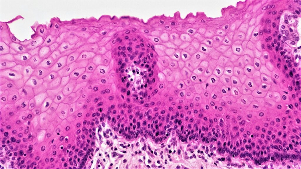
The wound healing process is how the body closes a breach in its skin: four overlapping phases — hemostasis, inflammation, proliferation and remodelling — each run by different cells on a different clock. In adult skin it ends in repair rather than regeneration, which is why a closed cut leaves a scar that never regains the full strength of the skin around it.
Below: each phase cell by cell, why chronic wounds stall, how tissue repair is studied, and what peptide research has and has not shown.
The short answer
- Four overlapping phases. Hemostasis (minutes to hours), inflammation (about days 1–3), proliferation (days 4–21) and remodelling (day 21 to a year or more).
- Platelets start the signalling. They adhere to the damaged vessel within seconds, and their granules release PDGF, TGF-β and VEGF.
- Neutrophils first, then macrophages, which shift from a pro-inflammatory toward a pro-repair state.
- Granulation tissue fills the gap: fibroblasts, capillaries and macrophages in a fibronectin and type III collagen matrix, re-covered by keratinocytes and pulled closed by myofibroblasts.
- Scars stay weaker. Type I collagen replaces type III, yet healed skin reaches at most about 80% of its original tensile strength.
- Chronic wounds are stalled repair. Diabetic, venous and pressure ulcers tend to stay locked in inflammation, with excess proteases.
- Peptide evidence is thin. LL-37 has two venous leg ulcer trials with mixed results; thymosin beta-4, BPC-157 and GHK-Cu rest mainly on cell and rodent work.
The wound healing process
Cutaneous wound repair · tissue repair · the stages of wound healing
- Phases
- Hemostasis, inflammation, proliferation, remodelling (overlapping)
- Approximate timing
- Minutes to hours; days 1–3; days 4–21; day 21 to a year or more
- How the gap closes
- Granulation tissue, keratinocyte migration and myofibroblast contraction
- Adult outcome
- A scar with about 80% of normal tensile strength
- Chronic wound
- No orderly closure within about three months
Where the peptide evidence stands
- Cell and ex vivo skin models
- Rodent wound models
- Small human trials (LL-37, thymosin beta-4)
- A research peptide established for wound closure
The phases are textbook biology; human data for catalogue peptides are a few small, mixed trials.
What the wound healing process is
The wound healing process is the coordinated cellular response that restores the skin barrier after injury. Guo and DiPietro describe four programmed phases of wound healing — hemostasis, inflammation, proliferation and remodelling — that must occur in the proper sequence and time frame for a wound to close well. Textbooks call them the stages of wound healing, but Landén, Li and Ståhle stress that the phases are sequential yet overlapping: cells of one phase are still at work when the next begins.
In adults the end product is usually a fibrotic scar rather than restored skin, whereas early-gestation fetal tissue can regenerate without scarring (Gurtner and colleagues, 2008).
Primary versus secondary intention
When a wound’s edges are closed directly, as in a sutured incision, it heals by primary intention. A wound left open heals by secondary intention, “from the base upwards, without direct closure”: granulation first, then re-epithelialization and contraction (Potluru and colleagues, 2025). Secondary intention relies far more on granulation tissue and contraction.
Hemostasis: the first minutes
Hemostasis (US: hemostasis) begins the moment a vessel is cut. Exposed collagen sets off the clotting cascades, and aggregating platelets trigger vasoconstriction (Landén). Platelets adhere to the injured vessel wall within seconds, the receptor GPIIb/IIIa cross-links them, and fibrin is deposited into the growing thrombus (Eisinger and colleagues, 2018). The clot also serves as a provisional matrix, a scaffold for cells that migrate in later.
Platelets also send the first repair signals. Their alpha granules release TGF-β1; PDGF-BB, a strong mitogen and chemoattractant for mesenchymal cells; and VEGF, which drives endothelial sprouting (Eisinger). PDGF and TGF-β from platelets, and later from macrophages, draw fibroblasts into the wound (Landén), and platelets release thymosin beta-4 at sites of injury (Treadwell and colleagues, 2012).
Inflammation and the macrophage switch
Inflammation starts within hours and dominates roughly the first three days. Neutrophils arrive first, recruited by damage- and pathogen-associated molecular patterns (Krzyszczyk and colleagues, 2018), and in an uninfected wound usually stay two to five days (Landén). Monocytes follow and become macrophages: one review places their arrival at about day 3 (Landén), while in mice they are recruited from about 24 hours after wounding (Krzyszczyk).
Early macrophages are mainly pro-inflammatory, releasing TNF-α, IL-1β, IL-6, reactive oxygen species and nitric oxide (see our NF-κB pathway explainer). Later ones secrete IL-10, TGF-β, PDGF, IGF-1 and VEGF. Clearing apoptotic neutrophils, called efferocytosis, helps drive the switch (Landén). The M1/M2 labels are, in one review’s words, “grossly oversimplified”: wound macrophages form a spectrum (Krzyszczyk). When the transition stalls, inflammation persists.
Proliferation and granulation tissue
From about day 4 to day 21, new connective tissue fills the defect, vessels grow into it and the epidermis closes over it (Landén).
What granulation tissue is made of
Granulation tissue is the temporary, vascular connective tissue that fills a wound from its base. Fibroblasts migrate in from the dermis, proliferate and deposit collagen, fibronectin, proteoglycans and hyaluronic acid; with new vessels and macrophages, that matrix is granulation tissue. The first collagen is largely type III, made quickly but weaker than the type I that later replaces it, and fibroblasts also secrete matrix metalloproteinases (MMPs) that degrade the provisional clot matrix (Landén). VEGF from platelets and macrophages drives the capillary sprouting covered in our angiogenesis and VEGF guide.
Re-epithelialization
Keratinocytes at the wound edge start migrating within hours to a day, and from about day 2 or 3 basal keratinocytes and nearby stem cells proliferate behind them (Landén). In human skin, re-epithelialization and granulation tissue are what drive closure (Galiano and colleagues, 2004).
Contraction and the myofibroblast
Some fibroblasts become myofibroblasts under mechanical tension and TGF-β; they express alpha-smooth muscle actin (α-SMA), pull the edges together and undergo apoptosis once repair is complete (Landén). Gabbiani, Ryan and Majno first described them at the University of Geneva in 1971 as “modified fibroblasts” in granulation tissue. A 2012 review led by Boris Hinz of the University of Toronto’s Faculty of Dentistry, with Gabbiani as senior author, covers how α-SMA expression is controlled and the cells’ role in fibrosis.
The remodelling phase and the scar
The remodelling phase begins as granulation tissue formation ends and can continue for a year or more. Type III collagen is gradually replaced by type I, which is deposited more slowly but is stronger, and vessel numbers fall (Landén). Myofibroblasts and vascular cells are removed by apoptosis, leaving a collagen-rich scar, and MMPs from fibroblasts and macrophages degrade matrix throughout (Krzyszczyk; Landén). Healed skin reaches a maximum of roughly 80% of its original tensile strength, and after serious injury hair follicles and sweat glands may not regenerate (Landén).
| Phase | Timing | Key cells | Key mediators and matrix | What a stained section shows |
|---|---|---|---|---|
| Hemostasis | Seconds to hours | Platelets | Fibrin; PDGF, TGF-β, VEGF | Aggregated platelets and fibrin filling the defect |
| Inflammation | Days 1–3 (neutrophils 2–5 days) | Neutrophils, then macrophages | TNF-α, IL-1β, IL-6; later IL-10, TGF-β | A neutrophil-rich infiltrate, increasingly replaced by macrophages |
| Proliferation | Days 4–21 | Fibroblasts, endothelial cells, keratinocytes, myofibroblasts | PDGF, TGF-β, VEGF; fibronectin, type III collagen | Granulation tissue (capillaries, fibroblasts, macrophages) under a thin advancing epidermis |
| Remodelling | Day 21 to a year or more | Fibroblasts | MMPs; type I replacing type III collagen | Fewer cells and vessels, denser collagen; hair follicles and glands may be absent |
Scarring versus regeneration
Adult mammalian skin repairs; some tissues and organisms regenerate, restoring the original architecture (Gurtner).
- Fetal skin. Human fetal skin from 15 to 22 weeks’ gestation, grafted onto athymic mice, repaired incisions without a scar when placed beneath the host’s skin but scarred when exposed to air; scarless repair needed neither fetal serum nor amniotic fluid (Lorenz and colleagues, 1992).
- Spiny mice. African spiny mice (Acomys) regenerate hair follicles in back-skin wounds, and their ear holes regenerate completely, with follicles, sebaceous glands, dermis and cartilage (Seifert and colleagues, Nature, 2012).
- Axolotls. Adult axolotls close full-thickness skin wounds without a scar, with a limited clot, fewer neutrophils, far fewer α-SMA-positive myofibroblasts than mouse wounds, little fibronectin and abundant tenascin-C; the authors present these differences as hypotheses to test (Seifert and colleagues, PLoS One, 2012).
Chronic wounds: repair that stalls
A chronic wound fails to progress through orderly repair to durable closure within an appropriate time, usually taken as about three months (Zhao and colleagues, 2016). It is the normal process stuck, most often in a self-perpetuating inflammatory stage: neutrophils release reactive oxygen species and proteases such as elastase and MMP-8, elastase degrades PDGF and TGF-β, and many wounds carry polymicrobial biofilms (Zhao). Macrophages stay pro-inflammatory, and chronic wound fluid has shown TNF-α and IL-1β about 100-fold higher than acute wound fluid (Krzyszczyk).
- Diabetic foot ulcers. Neuropathy, ischemia and trauma, plus glycation end-products, low-grade inflammation and microcirculatory dysfunction (Zhao); high glucose also impairs macrophage clearance of dead cells (Krzyszczyk).
- Venous leg ulcers. Venous hypertension lets macromolecules and red cells leak into tissue and attract leukocytes; these ulcers make up more than half of lower-limb chronic wounds (Zhao). Iron-overloaded, pro-inflammatory macrophages have been found in them (Landén).
- Pressure injuries. Sustained pressure or shear over bony prominences such as the sacrum, hips and malleoli causes ischemia; tissue dies from hypoxia and ischemia-reperfusion injury, sometimes after as little as two hours of immobility (Zhao).
Factors that slow repair
Guo and DiPietro list oxygenation, infection, age, sex hormones, stress, diabetes, obesity, medications, alcohol, smoking and nutrition as factors that can disrupt one or more phases; a bacterial burden above about 105 organisms per gram of tissue is linked to impaired repair (Zhao).
Physiology, not advice. This section explains how stalled repair is studied. It is not wound-care guidance.
How tissue repair is studied
Every model captures part of the wound healing process and misses the rest.
| Model | What it captures | Main limitation |
|---|---|---|
| Open excisional wound, rat or mouse | The whole sequence, cheaply | Closes largely by contraction; human wounds close mainly by re-epithelialization and granulation |
| Splinted excisional wound, mouse | A fixed ring blocks contraction, so closure runs through granulation and re-epithelialization over 2–4 weeks | Still mouse tissue; splinting itself increased granulation tissue |
| In-vitro scratch assay | Migration into a scratched gap, imaged over hours to overnight (Liang and colleagues, 2007) | One cell population on plastic |
| Ex vivo human skin wound | Re-epithelialization in organ-cultured human skin | Non-inflammatory, with no circulation |
Geoffrey Gurtner’s group described splinting in 2004, noting that no rodent model closely parallels human healing (Galiano); a 2013 methods paper co-written by Edward Tredget of the University of Alberta’s Wound Healing Research Group in Edmonton sets it out step by step (Wang and colleagues). In any peptide experiment, matched controls separate sequence-specific from non-specific effects (see scrambled peptide controls), and a conclusion’s weight depends on whether it comes from cells, animals or people (see how to read a peptide study).
LL-37, thymosin beta-4 and other repair-research peptides are listed in our recovery category, priced in CAD for laboratory research.
Browse recovery research peptidesPeptides in wound-repair research
Each peptide below is labelled by the strongest evidence we could confirm.
LL-37: small human trials, mixed results
LL-37, the C-terminal fragment of the human cathelicidin hCAP18, is better known as an antimicrobial peptide. Heilborn and colleagues (2003) found hCAP18 upregulated after skin injury, peaking around 48 hours, in the inflammatory infiltrate and migrating epithelium, but absent from the epithelium at chronic-ulcer edges; in organ-cultured human skin wounds, anti-LL-37 antibodies inhibited re-epithelialization in a concentration-dependent way.
A first double-blind trial in 34 patients with hard-to-heal venous leg ulcers and three concentration arms reported a healing-rate constant about six times placebo’s in the lowest arm (p = 0.003); the middle arm’s difference was not significant and the highest arm did not differ (Grönberg and colleagues, 2014). The phase IIb HEAL LL-37 trial in 148 patients found no significant difference from placebo overall; a post hoc large-wound subgroup differed, which the authors say needs an adequately powered trial (Mahlapuu and colleagues, 2021). LL-37 was well tolerated in both (see our LL-37 research guide).
Thymosin beta-4: rodent data, small human trials
In a 1999 rat full-thickness wound study, thymosin beta-4 increased re-epithelialization by 42% over saline at day 4 and up to 61% at day 7, with more contraction, collagen and angiogenesis (Malinda and colleagues). A 2012 review reports faster punch-wound closure in several rodent models and refers to two phase 2 trials, in venous stasis and pressure ulcers, without patient numbers or significance tests in its abstract (Treadwell). See thymosin alpha-1 vs thymosin beta-4 and BPC-157 vs TB-500.
BPC-157: rodent studies, almost no human data
Much of BPC-157’s wound literature comes from one University of Zagreb group, whose 2021 review covers skin wounds, burns, diabetic ulcers, fistulas and musculoskeletal injuries, mostly in rats and mice. It mentions earlier ulcerative colitis and multiple sclerosis trials without results and describes no human wound trials (Seiwerth and colleagues). Our BPC-157 safety review covers the human data.
GHK-Cu: cell and rodent work
In a 1993 rat study, implanted wound chambers that received the tripeptide–copper complex GHK-Cu accumulated more collagen, glycosaminoglycans, DNA and protein than saline chambers, in a concentration-dependent way, while a control tripeptide had no significant effect (Maquart and colleagues). That is matrix accumulation in a rodent chamber, not human wound closure; see our GHK-Cu research overview.
Becaplermin: the approved growth-factor example
Becaplermin, a form of PDGF, was in 1997 the first growth factor approved by the US FDA for a wound indication (diabetic foot ulcers); one review calls its benefit modest (Zhao). It is not a research peptide.
What’s established and what isn’t
Reasonably well established
- Skin repair runs through four overlapping phases, opened by platelet growth factors.
- Neutrophils arrive first; macrophages follow and shift toward a reparative state.
- Granulation tissue fills the gap, myofibroblasts contract it and keratinocytes re-cover it.
- Healed skin reaches about 80% of its original tensile strength.
- Chronic wounds show persistent inflammation and excess proteases.
Open questions
- Why fetal skin, axolotls and spiny mice repair without scars, and whether adult skin can be pushed that way.
- How well the M1/M2 labels describe wound macrophages.
- Whether LL-37 changes ulcer closure; the phase IIb trial found no overall difference.
- Whether rodent results for thymosin beta-4, BPC-157 and GHK-Cu translate to human skin.
Sourcing repair-research peptides in Canada
For wound-model work, the usual documentation questions apply, plus two that matter more in repair biology.
- Ask for a batch-matched certificate with HPLC purity and a mass-spectrometry identity check, and match its batch number to the vial (see our peptide COA guide).
- Ask about endotoxin, because wound models lean on inflammatory and macrophage readouts (see endotoxin in research peptides).
- Know which thymosin molecule you have. TB-500 and TB-500 Fragment are separate products.
- Store peptides cold, dry and dark, as in our peptide storage guide, and see our overview of research peptides in Canada for judging a supplier.
 LL-37Cathelicidin fragment studied in re-epithelialization and ulcer trialsView product
 TB-500 (Thymosin Beta-4)Thymosin beta-4; dermal wound data mainly from rodentsView product
 BPC-157Pentadecapeptide with wound studies mostly in rodentsView product
Research use only. Pure Peptides Canada products are laboratory reagents for in-vitro research, not for human or veterinary use. This article explains published research for education; it is not medical, dietary or dosing guidance.
Frequently asked questions
What are the four stages of wound healing?
The four stages, or phases, of wound healing are hemostasis, inflammation, proliferation and remodelling. Hemostasis forms a clot of platelets and fibrin, inflammation brings in neutrophils and then macrophages, proliferation builds granulation tissue and new epidermis, and remodelling matures the scar. The phases overlap.
How long does the wound healing process take?
One review gives approximate windows of minutes to hours for hemostasis, days 1 to 3 for inflammation, days 4 to 21 for proliferation, and day 21 to a year or more for remodelling. A wound can close within weeks while its scar keeps remodelling for months.
What is granulation tissue?
Granulation tissue is the temporary connective tissue that fills a wound from its base during the proliferation phase. It consists of fibroblasts, new capillaries and macrophages in a matrix of fibronectin, hyaluronic acid and collagen, mostly type III at first. Keratinocytes migrate over it, and during remodelling it matures into scar.
Why do some wounds become chronic?
Chronic wounds are usually stuck in inflammation. Excess neutrophils release proteases that degrade growth factors such as PDGF and TGF-beta, macrophages fail to switch to a reparative state, and many wounds carry biofilms. Diabetic, venous and pressure ulcers add neuropathy and ischemia, venous hypertension, or sustained pressure and shear.
What does peptide research show about wound healing?
The evidence is mostly preclinical. LL-37 has two placebo-controlled venous leg ulcer trials: the first reported a difference in its lowest-concentration arm, but the larger phase IIb trial found no significant difference overall. Thymosin beta-4, BPC-157 and GHK-Cu have been studied mainly in cells and rodents, and none is established to change wound closure in people.
Repair-research peptides for Canadian labs
LL-37, thymosin beta-4 and the rest of the recovery range are priced in Canadian dollars and shipped tracked from inside Canada. Checkout accepts credit and debit cards, Interac e-Transfer and cryptocurrency.
References
- Gabbiani G, Ryan GB, Majno G. Presence of modified fibroblasts in granulation tissue and their possible role in wound contraction. Experientia. 1971;27(5):549–550. DOI
- Lorenz HP, Longaker MT, Perkocha LA, Jennings RW, Harrison MR, Adzick NS. Scarless wound repair: a human fetal skin model. Development. 1992;114(1):253–259. DOI
- Maquart FX, Bellon G, Chaqour B, et al. In vivo stimulation of connective tissue accumulation by the tripeptide-copper complex glycyl-L-histidyl-L-lysine-Cu2+ in rat experimental wounds. J Clin Invest. 1993;92(5):2368–2376. DOI
- Malinda KM, Sidhu GS, Mani H, et al. Thymosin β4 accelerates wound healing. J Invest Dermatol. 1999;113(3):364–368. DOI
- Heilborn JD, Nilsson MF, et al. The cathelicidin anti-microbial peptide LL-37 is involved in re-epithelialization of human skin wounds and is lacking in chronic ulcer epithelium. J Invest Dermatol. 2003;120(3):379–389. DOI
- Galiano RD, Michaels VJ, Dobryansky M, Levine JP, Gurtner GC. Quantitative and reproducible murine model of excisional wound healing. Wound Repair Regen. 2004;12(4):485–492. DOI
- Liang CC, Park AY, Guan JL. In vitro scratch assay: a convenient and inexpensive method for analysis of cell migration in vitro. Nat Protoc. 2007;2(2):329–333. DOI
- Gurtner GC, Werner S, Barrandon Y, Longaker MT. Wound repair and regeneration. Nature. 2008;453:314–321. DOI
- Guo S, DiPietro LA. Factors affecting wound healing. J Dent Res. 2010;89(3):219–229. DOI
- Hinz B, Phan SH, Thannickal VJ, et al. Recent developments in myofibroblast biology. Am J Pathol. 2012;180(4):1340–1355. DOI
- Seifert AW, Kiama SG, Seifert MG, Goheen JR, Palmer TM, Maden M. Skin shedding and tissue regeneration in African spiny mice (Acomys). Nature. 2012;489:561–565. DOI
- Seifert AW, Monaghan JR, Voss SR, Maden M. Skin regeneration in adult axolotls: a blueprint for scar-free healing in vertebrates. PLoS One. 2012;7(4):e32875. DOI
- Treadwell T, Kleinman HK, Crockford D, Hardy MA, Guarnera GT, Goldstein AL. The regenerative peptide thymosin β4 accelerates the rate of dermal healing in preclinical animal models and in patients. Ann N Y Acad Sci. 2012;1270(1):37–44. DOI
- Wang X, Ge J, Tredget EE, Wu Y. The mouse excisional wound splinting model, including applications for stem cell transplantation. Nat Protoc. 2013;8(2):302–309. DOI
- Grönberg A, Mahlapuu M, Ståhle M, Whately-Smith C, Rollman O. Treatment with LL-37 is safe and effective in enhancing healing of hard-to-heal venous leg ulcers: a randomized, placebo-controlled clinical trial. Wound Repair Regen. 2014;22(5):613–621. DOI
- Landén NX, Li D, Ståhle M. Transition from inflammation to proliferation: a critical step during wound healing. Cell Mol Life Sci. 2016;73(20):3861–3885. DOI
- Zhao R, Liang H, Clarke E, Jackson C, Xue M. Inflammation in chronic wounds. Int J Mol Sci. 2016;17(12):2085. DOI
- Eisinger F, Patzelt J, Langer HF. The platelet response to tissue injury. Front Med. 2018;5:317. DOI
- Krzyszczyk P, Schloss R, Palmer A, Berthiaume F. The role of macrophages in acute and chronic wound healing and interventions to promote pro-wound healing phenotypes. Front Physiol. 2018;9:419. DOI
- Mahlapuu M, Sidorowicz A, Mikosinski J, et al. Evaluation of LL-37 in healing of hard-to-heal venous leg ulcers: a multicentric prospective randomized placebo-controlled clinical trial. Wound Repair Regen. 2021;29(6):938–950. DOI
- Seiwerth S, Milavic M, Vukojevic J, et al. Stable gastric pentadecapeptide BPC 157 and wound healing. Front Pharmacol. 2021;12:627533. DOI
- Potluru A, Pawlik O, Barlow R, Veitch D, Wernham AGH. A review of secondary intention healing in dermatology and dermatological surgery: part 1. Clin Exp Dermatol. 2025;50(6):1094–1100. DOI
For laboratory research use only. Not for human or veterinary use. This article summarizes published research for educational purposes and is not medical advice.