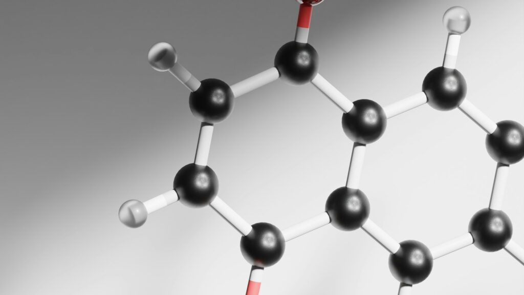
What is glutathione? It is a three-amino-acid peptide, γ-L-glutamyl-L-cysteinyl-glycine, that animal cells build for themselves and keep at millimolar concentrations, making it the most abundant low-molecular-weight thiol they produce. Its cysteine side chain gives cells a reusable supply of electrons for reducing peroxides, tagging reactive chemicals and shielding protein cysteines. For a laboratory it is also a reagent, and one that is easy to mismeasure.
This guide covers its structure, history, synthesis, functions and measurement, what the oral and liposomal glutathione trials measured, and its use as a reagent.
The short answer
- Glutathione is a tripeptide. γ-L-Glutamyl-L-cysteinyl-glycine links glutamate to cysteine through glutamate’s side-chain γ-carboxyl instead of the usual α-carboxyl, a structure that resists proteolysis. Its cysteine thiol does the chemistry: reduced glutathione (GSH) carries a free –SH, and two oxidized molecules join as glutathione disulfide (GSSG).
- It is abundant inside cells and scarce outside them. Mammalian cells hold about 0.5–10 mM; plasma typically about 2–3 µM.
- Two enzymes build it. Glutamate-cysteine ligase, the rate-limiting enzyme, is inhibited by glutathione and paced by cysteine supply; glutathione synthetase then adds glycine.
- Its jobs run through enzymes. Glutathione peroxidases use it to reduce peroxides, glutathione reductase regenerates it with NADPH, glutathione S-transferases conjugate it to electrophiles, and glutaredoxins remove it from proteins.
- The supplement trials measured levels, not outcomes. The oral and liposomal glutathione trials reviewed here were small, with mixed results, and none tested a clinical outcome.
- In the lab it is also a folding reagent. GSH/GSSG mixtures act as redox buffers for disulfide-containing proteins and peptides, and GSH oxidizes in solution.
Glutathione
GSH · γ-Glu-Cys-Gly · oxidized form: GSSG
- Full name
- γ-L-Glutamyl-L-cysteinyl-glycine
- Formula and mass
- C10H17N3O6S, about 307.3 g/mol (our calculation)
- Concentration
- About 0.5–10 mM in mammalian cells; about 2–3 µM in plasma
- Made by
- Glutamate-cysteine ligase (rate-limiting), then glutathione synthetase
- Recycled by
- Glutathione reductase, using NADPH
- Broken down by
- γ-Glutamyl transferase (GGT), outside the cell
- First isolated
- 1921, by Frederick Gowland Hopkins; shown to be a tripeptide in 1929
Where the evidence stands
- Structure and enzymology
- Cell and animal models
- Small human biomarker trials
- Clinical outcomes from supplementation
The biochemistry is textbook material. None of the supplement trials reviewed here tested a clinical outcome.
What is glutathione?
Glutathione is a tripeptide of glutamate, cysteine and glycine with one unusual bond. In an ordinary peptide, each bond is made through an amino acid’s α-carboxyl; in glutathione, glutamate is linked to cysteine through the γ-carboxyl at the end of its side chain. Aquilano and colleagues note that this structure makes glutathione resistant to proteolysis, with breakdown initiated by γ-glutamyl transferase (GGT). See our guides to dipeptides, tripeptides and oligopeptides and amino acid codes.
The working part is cysteine’s thiol (–SH). In reduced glutathione (GSH) it is free. When two GSH molecules each give up an electron and a proton, their sulfur atoms join as glutathione disulfide (GSSG), and enzymes convert GSSG back again.
Forman and colleagues describe glutathione as the most abundant low-molecular-weight thiol compound synthesized in cells. Mammalian cells contain 0.5–10 mM depending on the tissue, with the liver at about 5–10 mM (Aoyama), whereas more than 98% of blood glutathione is inside red cells and plasma GSH is typically 2–3 µM (Giustarini and colleagues, 2026). In the cells Hwang and colleagues studied, the overall GSH:GSSG ratio was 30:1 to 100:1.
How glutathione was identified
In 1888 de Rey-Pailhade described a substance that reduced sulfur to hydrogen sulfide and called it “philothion”. In 1921 Frederick Gowland Hopkins isolated what he took to be a dipeptide of glutamate and cysteine from yeast and animal tissues, in a paper titled “On an autoxidisable constituent of the cell”. In 1929 Hopkins showed it was a tripeptide that also contains glycine; that year Edward C. Kendall crystallized it and identified its structure (Aoyama).
Glutathione synthesis: two enzymes and a bottleneck
Glutathione synthesis takes place mainly in the cytosol, in two energy-requiring steps (Orlowski and Meister; Aquilano and colleagues). Glutamate-cysteine ligase (GCL), historically called γ-glutamylcysteine synthetase, joins glutamate’s γ-carboxyl to cysteine to make γ-glutamylcysteine; glutathione synthetase then adds glycine. GCL has a catalytic (GCLC) and a modifier (GCLM) subunit and is the rate-limiting enzyme (Lu, 2013). Three controls set its pace:
- Product feedback. Richman and Meister showed in 1975 that glutathione inhibits the enzyme under conditions similar to those inside cells, binding at its glutamate site rather than at an allosteric site.
- Cysteine supply. They measured an apparent Km for cysteine of 0.35 mM, close to intracellular cysteine levels, and inside cells cysteine is far scarcer than glutamate or glycine (Aoyama).
- Gene expression. In HepG2 cells, the transcription factor Nrf2 bound electrophile response elements in the promoters of both GCL subunit genes and raised their activity (Wild and colleagues); see Nrf2 and oxidative stress.
The loop also explains 5-oxoprolinuria: with glutathione synthetase markedly deficient, the unrestrained first enzyme overproduces γ-glutamylcysteine, which becomes 5-oxoproline faster than 5-oxoprolinase can convert it to glutamate (Richman and Meister).
Glutathione function: the main jobs
Most of what glutathione does, it does as a substrate for enzymes.
| Job | Enzyme or setting | What happens to GSH |
|---|---|---|
| Reducing peroxides | Glutathione peroxidases (selenium-dependent) | Two GSH become one GSSG |
| Recycling | Glutathione reductase, using NADPH | GSSG is reduced back to two GSH |
| Conjugating electrophiles | Glutathione S-transferases | GSH is consumed as a conjugate |
| Protecting protein thiols | S-glutathionylation, reversed by glutaredoxins | GSH is attached to a protein cysteine through a disulfide, then removed |
| Supplying cysteine | γ-Glutamyl transferase on the cell surface | Exported GSH is split; its cysteinylglycine can feed new synthesis |
Peroxides and the GSH/GSSG cycle
Selenium-dependent glutathione peroxidases are the main route by which glutathione reduces hydrogen peroxide and lipid hydroperoxides (Aquilano and colleagues). Two GSH become one GSSG, and glutathione reductase regenerates GSH using NADPH (Giustarini and colleagues, 2026), a phosphorylated relative of the coenzyme covered in our guide to NAD+, NMN and nicotinamide riboside. In a 2014 study that profiled 177 cancer cell lines and used mouse xenografts, Yang and colleagues identified GPX4 as an essential regulator of ferroptosis, a non-apoptotic cell death, and found that some ferroptosis inducers acted by depleting glutathione; these are cancer-model findings.
Conjugation: the NAPQI example
Glutathione S-transferases (GSTs) carry out most conjugation of glutathione with electrophiles, reactive electron-seeking molecules (Aquilano and colleagues). In mouse liver microsomes, Dahlin and colleagues showed in 1984 that cytochrome P-450 oxidizes acetaminophen (paracetamol) to the reactive metabolite NAPQI, which binds covalently to protein; glutathione reduced that binding and formed the conjugate 3-S-glutathionylacetaminophen. This is chemistry, not drug guidance.
S-glutathionylation and cysteine storage
In S-glutathionylation, glutathione is attached through a disulfide to a protein cysteine. Aquilano and colleagues describe the modification as protecting cysteines from irreversible oxidation while relaying redox signals, and glutaredoxin reverses it. They also list storage of cysteine reserves among glutathione’s functions.
Compartments: mitochondria and the endoplasmic reticulum
The cytosol, the main site of synthesis, holds almost 90% of cellular glutathione, which is distributed from there to mitochondria, the nucleus and the endoplasmic reticulum (Aquilano and colleagues). Mitochondria therefore import it: in 2021 Wang and colleagues identified the carrier SLC25A39 as necessary for that import in mammalian cells, and in mice the import was needed for red blood cell development. The secretory pathway is the opposite case: Hwang and colleagues found glutathione to be its major redox buffer, at a GSH:GSSG ratio of only 1:1 to 3:1.
Breakdown: GGT and the γ-glutamyl cycle
Breakdown begins outside the cell with GGT, an enzyme on the cell surface, which removes the γ-glutamyl group and leaves cysteinylglycine; a peptidase then releases cysteine and glycine, which cells such as neurons take up for new synthesis (Aquilano and colleagues; Aoyama). Working with rat kidney enzymes in 1970, Orlowski and Meister named this loop the γ-glutamyl cycle and proposed it as an amino acid transport system.
Glutathione (GSH) is listed with our cellular-research reagents, priced in CAD.
Browse anti-aging & cellular researchHow laboratories measure glutathione
Three families of method dominate, and all share one weakness: GSH oxidizes easily, so careless handling manufactures GSSG.
- Ellman’s reagent. DTNB reacts with GSH to release a product that absorbs strongly at 412 nm (Giustarini and colleagues, 2026), but it reacts with any soluble thiol (Tomin and colleagues).
- Enzymatic recycling. Glutathione reductase and NADPH keep turning GSSG back into GSH, which reacts with DTNB again (Giustarini and colleagues, 2026); Tietze’s 1969 method used this principle to measure total and oxidized glutathione.
- Chromatography and mass spectrometry. HPLC with fluorescence detection can measure GSH, and LC-MS/MS can quantify GSH and GSSG without enzyme coupling (Giustarini and colleagues, 2026; Tomin and colleagues). See our guides to HPLC testing and LC-MS.
Acid deproteinization can oxidize GSH and inflate GSSG, so careful methods block the thiol with N-ethylmaleimide (NEM) first (Giustarini and colleagues, 2026). Our article on GSH vs GSSG works through the size of the oxidation artifact.
Oral and liposomal glutathione: what trials measured
Glutathione is sold in plain oral and liposomal forms. The controlled trials measured glutathione levels and laboratory markers, not health outcomes; we report them as research, not dietary guidance.
The absorption problem
In 1992 Witschi and colleagues found no significant rise in plasma glutathione, cysteine or glutamate over 270 minutes after a single oral intake in seven healthy volunteers, and attributed the negligible systemic availability to hydrolysis by intestinal and hepatic GGT. In 2011 a randomized, double-blind, placebo-controlled trial by Allen and Bradley in 40 adult volunteers (39 completed) found no significant change after four weeks in two urinary oxidative-stress markers or in red-cell GSH, GSSG or their ratio.
Trials that reported higher levels
Richie and colleagues randomized 54 non-smoking adults to placebo or to oral glutathione at two intake levels for six months, double-blind. In the higher-intake arm, glutathione rose by roughly 30–35% in red cells, plasma and lymphocytes and by about 260% in buccal cells; increases were generally larger at the higher intake and with longer use, and levels returned to baseline after a one-month washout.
Liposomal products enclose glutathione in lipid vesicles (liposomes). In a one-month pilot study published in 2018, Sinha and colleagues gave liposomal glutathione at two intake levels to 12 healthy adults; the abstract describes no placebo group. Glutathione peaked at week two about 40% above baseline in whole blood and about 100% in peripheral blood mononuclear cells, with no difference between intake groups, and the authors called their findings preliminary.
What these trials cannot show. The four studies enrolled 7 to 54 volunteers for anything from a single sitting to six months and measured biomarkers, not clinical outcomes. The two positive trials share several authors and did not compare liposomal with plain glutathione head to head. See our guide to reading a peptide study.
Skin-lightening marketing and regulator warnings
Glutathione is also heavily marketed for skin lightening, on mechanisms proposed from pigment biochemistry: tyrosinase inhibition and a shift from dark eumelanin toward lighter pheomelanin. A 2016 review by Sonthalia and colleagues found only three randomized trials, of oral or topical forms, and called for larger, well-designed trials with long-term follow-up. It found no evidence of efficacy for intravenous glutathione, the subject of a 2019 warning: the Food and Drug Administration of the Philippines’ Advisory No. 2019-182, “Unsafe use of glutathione as skin lightening agent”, addressed drips offered by salons, spas and beauty clinics, said the agency had approved no such product for skin lightening, and listed possible toxic effects on the liver, kidneys and nervous system, possible Stevens-Johnson syndrome and infection risks. We make no recommendation about any cosmetic use.
Glutathione as a laboratory reagent
In the laboratory, glutathione is commonly used to help disulfide-containing proteins and peptides form correct disulfide bonds. A GSH/GSSG mixture acts as a redox buffer whose thiols undergo reversible thiol-disulfide exchange with the unfolded chain, the same chemistry Hwang and colleagues observed in the secretory pathway.
In 1975 Ahmed, Schaffer and Wetlaufer reactivated reduced bovine pancreatic ribonuclease in glutathione buffers in yields above 90%, and the recovered enzyme matched native ribonuclease in specific activity, chromatographic behaviour and circular dichroism. Their optimum, roughly ten parts GSH to one part GSSG at millimolar GSH, matched earlier lysozyme work. For synthetic cystine-knot peptides, Reinwarth and colleagues list high dilution and redox folding assistants such as oxidized and reduced glutathione among the common requirements.
Glutathione oxidizes in solution. The OIV specification for glutathione used in winemaking stores the solid in sealed packaging in a cool (2–8 °C), dry place, has its GSH test solution prepared each day and kept cold in a brown glass bottle, and calls for precautions against oxidation to GSSG. A buffer keeps its weighed-out GSH:GSSG ratio only while it is fresh. See our peptide storage guide and how peptides degrade.
What’s established and what isn’t
Reasonably well established
- Glutathione is γ-glutamyl-cysteinyl-glycine; it resists proteolysis, and GGT initiates its breakdown.
- GCL (rate-limiting, inhibited by glutathione) and glutathione synthetase make it, mainly in the cytosol.
- Cysteine supply limits synthesis, and Nrf2 drives both GCL subunit genes (shown in HepG2 cells).
- Glutathione peroxidases, glutathione reductase and GSTs use, recycle and conjugate GSH.
- SLC25A39 is needed for mitochondrial glutathione import (cells and mice).
- A single oral intake did not raise plasma glutathione in volunteers.
Open questions
- Whether oral or liposomal glutathione changes any clinical outcome.
- Whether liposomal formulation adds anything over plain glutathione.
- Why six months of intake raised cellular levels in one trial when four weeks changed nothing in another.
- Whether glutathione affects skin pigmentation.
- Whether GPX4 and ferroptosis findings from cancer models apply to people.
Sourcing glutathione for research in Canada
For a Canadian lab buying glutathione as a reagent, the usual documentation questions apply, plus one specific to a thiol: how much is still reduced. Pure Peptides Canada is itself a supplier, so apply these checks to us too; our overview of research peptides in Canada explains how to verify a supplier.
- Confirm the form. By our calculation GSH averages about 307.3 g/mol and GSSG about 612.6 g/mol, so an identity result near 612–613 means the oxidized form (see our GSH vs GSSG guide).
- Ask about GSSG. It is the impurity that tracks oxidation. Our HPLC testing guide explains the method behind most purity results.
- Match the batch. The certificate’s batch number should match your vial; our peptide COA guide covers what else to check.
 Glutathione (GSH)Reduced glutathione, the γ-Glu-Cys-Gly tripeptide this guide describesView product
 NAD+A nicotinamide coenzyme; its phosphorylated relative NADPH supplies glutathione reductaseView product
 HumaninA mitochondrial peptide, for labs studying the organelle that must import glutathioneView product
Research use only. Pure Peptides Canada products are laboratory reagents for in-vitro research, not for human or veterinary use. This article explains published research for education; it is not medical, dietary or dosing guidance.
Frequently asked questions
What is glutathione?
Glutathione is a tripeptide, γ-L-glutamyl-L-cysteinyl-glycine, that animal cells make from glutamate, cysteine and glycine. It is the most abundant low-molecular-weight thiol that cells synthesize, at about 0.5–10 mM in mammalian cells. Enzymes use its cysteine thiol to reduce peroxides, conjugate reactive chemicals and modify protein cysteines.
Is glutathione a peptide or a protein?
It is a peptide, only three amino acids long. Its glutamate is linked to cysteine through the side-chain γ-carboxyl rather than the α-carboxyl of an ordinary peptide bond, which makes it resistant to proteolysis. Cells build it with two dedicated enzymes.
How does the body make glutathione?
In two steps, mainly in the cytosol. Glutamate-cysteine ligase, the rate-limiting enzyme, joins glutamate and cysteine into γ-glutamylcysteine, and glutathione synthetase adds glycine. Glutathione inhibits the first enzyme, cysteine supply limits it, and the transcription factor Nrf2 raises expression of its subunit genes.
What does glutathione do in cells?
Glutathione peroxidases use it to reduce peroxides, producing GSSG, which glutathione reductase converts back to GSH with NADPH. Glutathione S-transferases attach it to reactive electrophiles, and proteins are reversibly S-glutathionylated on their cysteines.
Is oral or liposomal glutathione absorbed?
The evidence is limited and mixed. A single oral intake did not raise plasma glutathione in a 1992 study, and a four-week placebo-controlled trial found no change. A six-month placebo-controlled trial and a one-month liposomal pilot reported higher levels in blood and cells, but both were small, measured only biomarkers and came from overlapping teams.
Does glutathione lighten skin?
It is heavily marketed for that, but the evidence is limited. A 2016 review found only three randomized trials of oral or topical forms and called for larger, well-designed studies. In 2019 the Food and Drug Administration of the Philippines issued an advisory titled “Unsafe use of glutathione as skin lightening agent”, warning about glutathione drips offered for that purpose.
Glutathione for redox research
Reduced glutathione is stocked as a research reagent alongside NAD+. Orders are priced in Canadian dollars, ship tracked from inside Canada by Canada Post Xpresspost, and can be paid by credit or debit card, Interac e-Transfer or cryptocurrency at checkout.
References
- Hopkins FG. On an autoxidisable constituent of the cell. Biochem J. 1921;15(2):286–305. DOI
- Tietze F. Enzymic method for quantitative determination of nanogram amounts of total and oxidized glutathione: applications to mammalian blood and other tissues. Anal Biochem. 1969;27(3):502–522. DOI
- Orlowski M, Meister A. The γ-glutamyl cycle: a possible transport system for amino acids. Proc Natl Acad Sci USA. 1970;67(3):1248–1255. DOI
- Richman PG, Meister A. Regulation of gamma-glutamyl-cysteine synthetase by nonallosteric feedback inhibition by glutathione. J Biol Chem. 1975;250(4):1422–1426. DOI
- Ahmed AK, Schaffer SW, Wetlaufer DB. Nonenzymic reactivation of reduced bovine pancreatic ribonuclease by air oxidation and by glutathione oxidoreduction buffers. J Biol Chem. 1975;250(21):8477–8482. DOI
- Dahlin DC, Miwa GT, Lu AYH, Nelson SD. N-acetyl-p-benzoquinone imine: a cytochrome P-450-mediated oxidation product of acetaminophen. Proc Natl Acad Sci USA. 1984;81(5):1327–1331. DOI
- Witschi A, Reddy S, Stofer B, Lauterburg BH. The systemic availability of oral glutathione. Eur J Clin Pharmacol. 1992;43(6):667–669. DOI
- Hwang C, Sinskey AJ, Lodish HF. Oxidized redox state of glutathione in the endoplasmic reticulum. Science. 1992;257(5076):1496–1502. DOI
- Wild AC, Moinova HR, Mulcahy RT. Regulation of γ-glutamylcysteine synthetase subunit gene expression by the transcription factor Nrf2. J Biol Chem. 1999;274(47):33627–33636. DOI
- Forman HJ, Zhang H, Rinna A. Glutathione: overview of its protective roles, measurement, and biosynthesis. Mol Aspects Med. 2009;30(1–2):1–12. DOI
- Allen J, Bradley RD. Effects of oral glutathione supplementation on systemic oxidative stress biomarkers in human volunteers. J Altern Complement Med. 2011;17(9):827–833. DOI
- Reinwarth M, Nasu D, Kolmar H, Avrutina O. Chemical synthesis, backbone cyclization and oxidative folding of cystine-knot peptides: promising scaffolds for applications in drug design. Molecules. 2012;17(11):12533–12552. DOI
- Lu SC. Glutathione synthesis. Biochim Biophys Acta. 2013;1830(5):3143–3153. DOI
- Aquilano K, Baldelli S, Ciriolo MR. Glutathione: new roles in redox signaling for an old antioxidant. Front Pharmacol. 2014;5:196. DOI
- Yang WS, SriRamaratnam R, Welsch ME, et al. Regulation of ferroptotic cancer cell death by GPX4. Cell. 2014;156(1–2):317–331. DOI
- Richie JP Jr, Nichenametla S, Neidig W, et al. Randomized controlled trial of oral glutathione supplementation on body stores of glutathione. Eur J Nutr. 2015;54(2):251–263. DOI
- Sonthalia S, Daulatabad D, Sarkar R. Glutathione as a skin whitening agent: facts, myths, evidence and controversies. Indian J Dermatol Venereol Leprol. 2016;82(3):262–272. DOI
- Sinha R, Sinha I, Calcagnotto A, et al. Oral supplementation with liposomal glutathione elevates body stores of glutathione and markers of immune function. Eur J Clin Nutr. 2018;72(1):105–111. DOI
- Food and Drug Administration of the Philippines. FDA Advisory No. 2019-182: Unsafe use of glutathione as skin lightening agent. July 5, 2019. FDA Philippines
- Tomin T, Schittmayer M, Birner-Gruenberger R. Addressing glutathione redox status in clinical samples by two-step alkylation with N-ethylmaleimide isotopologues. Metabolites. 2020;10(2):71. DOI
- Aoyama K. Glutathione in the brain. Int J Mol Sci. 2021;22(9):5010. DOI
- Wang Y, Yen FS, Zhu XG, et al. SLC25A39 is necessary for mitochondrial glutathione import in mammalian cells. Nature. 2021;599(7883):136–140. DOI
- Giustarini D, Colombo G, Dalle-Donne I, Rossi R. An artifact-free assay for the GSH/GSSG ratio adapted for finger-stick blood microvolumes: simple, sensitive, and suitable for any laboratory. Antioxidants. 2026;15(4):483. DOI
- International Organisation of Vine and Wine (OIV). COEI-1-GLUTAT: Glutathione. International Oenological Codex. OIV
For laboratory research use only. Not for human or veterinary use. This article summarizes published research for educational purposes and is not medical advice.