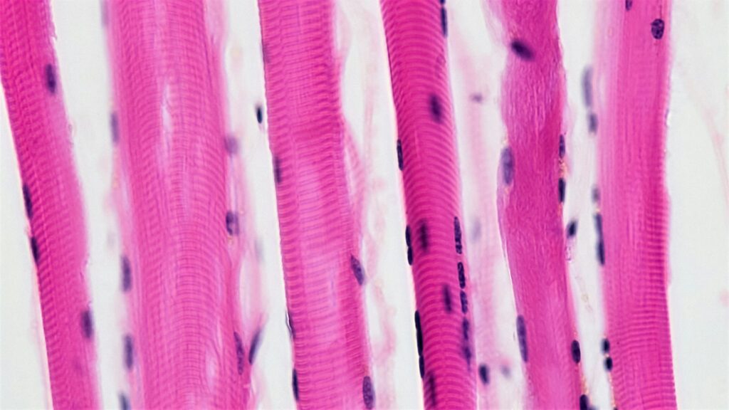
Satellite cells are the resident stem cells of skeletal muscle: small cells pressed against each muscle fibre, under the sheet of matrix that wraps it, that wake after injury, multiply and fuse to rebuild the fibre. Mouse muscle cannot regenerate after acute injury without them, and they sit at the centre of two live debates: whether fibres need them to enlarge, and whether the nuclei they donate leave a lasting “muscle memory”.
This guide covers their discovery and Canadian research history, their niche and states, the regeneration, hypertrophy and muscle-memory evidence, aging, and where research compounds fit.
The short answer
- They are muscle stem cells. Satellite cells sit between a muscle fibre’s plasma membrane (the sarcolemma) and its basal lamina, and make up roughly 5% of fibre-associated nuclei in adult rodents.
- Pax7 marks them. Quiescent satellite cells express Pax7 and need Notch signalling to stay at rest; activated cells switch on Myf5 and MyoD, and myogenin marks those that differentiate and fuse.
- They renew themselves. Cells that keep Pax7 high return to rest, and in mice an asymmetric division can give one stem-like and one committed daughter.
- Regeneration needs them. In three 2011 mouse studies, removing Pax7-expressing cells stopped injured muscle from regenerating.
- Hypertrophy is contested. Fibres in satellite-cell-depleted mice enlarged normally over two weeks of overload in one study but not in another lab’s repeat, and longer overload impaired growth.
- Muscle memory is unresolved. Rodent studies found extra myonuclei kept for months; a later mouse study and a human study did not confirm it, and a small human study points to marks in muscle DNA instead.
- Age wears them down. Older muscle has fewer satellite cells on type II fibres; in aged mice, niche FGF2 and senescence impair them, and the rescues cited here are mouse-only.
Satellite cells
muscle satellite cells · myosatellite cells · muscle stem cells (MuSCs)
- What they are
- Resident stem cells of skeletal muscle
- Where they sit
- Between the fibre’s plasma membrane and its basal lamina
- How common
- About a third of muscle nuclei around birth in rats; roughly 5% in adult rodents
- Defining marker
- Pax7, a paired-box transcription factor
- First described
- Alexander Mauro, 1961, by electron microscopy of frog muscle
- Canadian milestones
- Pax7 requirement (McMaster, 2000); asymmetric self-renewal (Ottawa, 2007)
Where the evidence stands
- Identified in tissue
- Required for regeneration in mice
- Counted in human biopsies
- A settled role in hypertrophy and muscle memory
Their role in repair is settled in mice; whether fibres need them to enlarge, and whether their nuclei store a memory of training, is still argued over.
What are satellite cells?
A satellite cell is a single-nucleus stem cell with very little cytoplasm, wedged between a skeletal muscle fibre’s plasma membrane, the sarcolemma, and the basal lamina, a thin sheet of extracellular matrix around the fibre. Muscle fibres are very large cells whose many nuclei, the myonuclei, share one cytoplasm; when a fibre gains nuclei during growth or repair, they come from satellite cells that fuse into it. In rats, satellite cells account for about a third of muscle nuclei around birth, about 10% at four weeks of age and around 5% in adults.
How researchers find them
Until molecular markers arrived, confirming a satellite cell’s identity required electron microscopy. Today tissue sections are stained for Pax7, standard in animals and increasingly in human biopsies, or NCAM (CD56), plus laminin to outline the basal lamina. A single marker can miss cells, so double labelling is preferred, and reliable fibre-type-specific counts in young men need at least 50 type I and 75 type II fibres. Choices like these explain part of why the studies below disagree.
Discovery, and the Canadian labs that defined the markers
In 1961 Alexander Mauro, examining frog tibialis anticus muscle by electron microscopy, described and named the satellite cell, reported similar cells in other frog muscles and in rat muscle, and proposed that they might be dormant myoblasts held in reserve to rebuild damaged fibres. Bernard Katz reported similar cells on frog muscle-spindle fibres the same month.
Two landmark marker studies came from Ontario. In 2000, Patrick Seale and colleagues in Michael Rudnicki’s group at McMaster University in Hamilton, with collaborators at the Max Planck Institute for Biophysical Chemistry in Göttingen, found no satellite cells in the muscle of mice lacking Pax7 and concluded that Pax7 is required to specify the lineage. Rudnicki’s lab later worked in Ottawa, at the Ottawa Health Research Institute (the Ottawa Hospital Research Institute in its later papers), where it mapped how satellite cells renew themselves. McMaster kinesiologist Gianni Parise co-authored two of the reviews cited here.
From quiescence to fusion: how satellite cells work
A satellite cell’s life is a loop: rest, activation, expansion, then differentiation and fusion or a return to rest.
- Quiescence. Notch signalling is active in resting satellite cells. When Thomas Rando’s group deleted RBP-Jκ, the nuclear factor Notch signalling requires, mouse satellite cells activated spontaneously, differentiated mostly without dividing and fused into fibres until the pool drained and muscle could no longer regenerate.
- Activation. Hepatocyte growth factor (HGF), acting through its receptor c-Met, is one activating signal. Myf5 is the earliest marker of commitment, followed by MyoD. In young men, satellite-cell numbers rose within 24 hours of a bout of eccentric exercise and peaked at around 72 hours, and most satellite cells stained for IGF-1 a day after such a bout, though not at rest; see our IGF-1 explainer.
- Differentiation and fusion. Cells that differentiate switch on myogenin and MRF4 and switch off Pax7, then fuse, building new fibres or adding myonuclei to existing ones.
- Self-renewal. Cells that keep Pax7 high return to quiescence. In 2007, Shihuan Kuang and colleagues in Rudnicki’s Ottawa lab found by lineage tracing in mice that about 10% of Pax7-positive satellite cells had never expressed Myf5. These divided along the apical-basal axis into a basal Pax7+/Myf5− daughter and an apical Pax7+/Myf5+ daughter; after transplantation, Pax7+/Myf5− cells spread through the host’s satellite-cell pool while Pax7+/Myf5+ cells differentiated prematurely. In 2015 the lab reported that dystrophin, the protein missing in Duchenne muscular dystrophy, is needed in activated satellite cells for polarity and asymmetric division, implicating faulty stem cells as well as fragile fibres in the disease.
Satellite cells and muscle regeneration: the 2011 ablation studies
Transplantation had shown that satellite cells can rebuild muscle, but other cell types had also been reported to contribute to fibres, so their necessity was untested. In 2011, three groups settled it in one issue of Development by genetically removing Pax7-expressing cells from mice:
- Lepper, Partridge and Fan found regenerative myogenesis completely blocked after tibialis anterior injury and in grafted muscles.
- Sambasivan and colleagues put a human diphtheria toxin receptor under the mouse Pax7 locus. After diphtheria toxin, muscle failed to regenerate and filled with inflammatory and fat cells; no other cell type compensated, and transplanting Pax7-positive satellite cells alone rescued regeneration.
- Murphy and colleagues in Gabrielle Kardon’s lab removed satellite cells or Tcf4-positive fibroblasts. Without satellite cells no regenerated muscle formed and connective tissue expanded; without fibroblasts, satellite cells differentiated too early and regenerated fibres were smaller.
Satellite cells are therefore indispensable for regeneration after acute injury in mice, with niche fibroblasts regulating their expansion; the inflammatory side of that response is covered in our guide to the overlapping phases of tissue repair.
One research peptide has been tested in regenerating dystrophic muscle: in mdx mice, a Duchenne model, six months of thymosin beta-4 (Tβ4) increased the number of regenerating fibres, but grip strength, cardiac function and fibrosis did not improve significantly. That is a single mouse study; for how full-length Tβ4 relates to the TB-500 names, see TB-500 vs TB-500 fragment.
Thymosin beta-4 (TB-500) and the rest of the recovery and repair range are laboratory reagents for in-vitro research, priced in CAD.
Browse recovery & repairDo satellite cells drive hypertrophy? The myonuclear domain debate
The myonuclear domain theory holds that each myonucleus supports a limited volume of cytoplasm, so fibre size and myonuclear number rise together: satellite cells donate nuclei during hypertrophy, and nuclei are lost by apoptosis during atrophy. Ablation tools let researchers test it.
- Two weeks without satellite cells. John McCarthy and Charlotte Peterson’s group removed over 90% of satellite cells from adult mice and overloaded the plantaris by surgically removing its synergists. After two weeks, fibres enlarged as much as in controls without adding myonuclei, so each nucleus served more cytoplasm.
- Eight weeks without them. Longer overload in the same model impaired hypertrophy, which the authors linked to increased extracellular matrix (Fry and colleagues, 2014, as summarized in a 2015 review).
- A repeat that disagreed. Kristian Gundersen’s Oslo group, using immunohistochemistry and in vivo imaging, found hypertrophy prevented in satellite-cell-deficient mice and attributed the earlier result to using muscle mass as a proxy for fibre size and to counting regenerating fibres.
In people, satellite-cell and myonuclear content rise alongside hypertrophy during prolonged resistance training, but that is correlation, not proof of necessity.
Why the results differ. The studies varied in overload duration, the muscles examined and whether fibre size was measured directly. A 2015 McMaster and Maastricht review concluded that satellite cells appear necessary for extensive hypertrophy, a conclusion drawn from mouse data only.
Muscle memory: myonuclei, epigenetics and the debate
Muscle memory usually means that previously trained muscle regains size and strength faster. Part of it is neural: in a 2019 human study, strength stayed about 60% above baseline after 20 weeks without training although muscle size had returned to baseline, which the authors read as lasting motor learning. The cellular question is whether fibres keep a record, and satellite cells matter because they supply new myonuclei.
The case for a myonuclear memory rests on two Oslo rodent studies; the doubts come from a Kentucky mouse study and a human study that could not test the idea (table below). A 2020 Maastricht and McMaster review concluded that most of the evidence comes from rodents and that whether it holds in people is unclear. A small 2018 human study proposes a different store: methylation marks in muscle DNA that persisted through a training break.
| Claim | Model | Finding | Caveat |
|---|---|---|---|
| Myonuclei survive atrophy (Oslo, 2010) | Rodents; in vivo imaging of single fibres | Overload added myonuclei before fibres enlarged much; they survived long denervation atrophy | Denervation is not ordinary detraining |
| Retained myonuclei speed regrowth (Oslo, 2013) | Female mice; 14-day testosterone propionate exposure | Myonuclei up 66% and still raised at least three months after withdrawal, though fibre size normalized within three weeks; later overload enlarged fibres 31% in six days, with no significant change in controls | Hormone, not exercise; same laboratory |
| Training-gained myonuclei are lost (Kentucky, 2019) | Female mice; 8 weeks of weighted wheel running, then 12 weeks off | Fewer myonuclei as the plantaris returned to an untrained state | Mainly fast-twitch muscle |
| A trained leg retrains faster (2019) | 19 adults; one leg trained, 20 weeks off, both legs retrained | The two legs responded alike | The first training block added no myonuclei, so the test was not rigorous |
| Muscle DNA keeps a memory (2018) | 8 untrained men; 7 weeks training, 7 off, 7 retraining | Lean mass up 6.5%, back to baseline, then up 12.4%; 18,816 hypomethylated sites after retraining vs 9,153 after training, with some genes hypomethylated through the break | Tiny all-male sample; whole-tissue DNA |
Physiology, not advice. These studies describe nuclei and DNA in specific models and are not a basis for training decisions.
Satellite cells in aging muscle
In older muscle, type II fibres are smaller and carry fewer satellite cells, and the satellite-cell response to exercise is blunted in older men. Mouse work points to three causes:
- The niche. Aged mouse fibres express FGF2, which drives a subset of satellite cells out of quiescence and erodes their self-renewal; inhibiting FGF receptor 1 signalling or overexpressing Sprouty1, an FGF-signalling inhibitor, prevented the depletion.
- The blood. Joining the circulations of young and old mice (heterochronic parabiosis) restored Notch activation, proliferation and regenerative capacity in aged satellite cells.
- The cells. In geriatric mice, resting satellite cells slid from reversible quiescence into a pre-senescent state through p16INK4a; silencing it restored regeneration, and human geriatric satellite cells also showed p16INK4a dysregulation. See our senolytics explainer.
Two metabolic routes have been tested. In a 2016 study from Johan Auwerx’s Lausanne group, with the University of Ottawa’s Keir Menzies as a co-corresponding author, the NAD+ precursor nicotinamide riboside restored muscle stem cell function in aged mice through the mitochondrial unfolded protein response and extended mouse lifespan (see NAD+, NMN and nicotinamide riboside). In 2019, a University of Texas Medical Branch group that develops inhibitors of nicotinamide N-methyltransferase (NNMT), an enzyme linked to impaired NAD+ salvage and dysregulated SIRT1 activity (sirtuins explained), gave injured 24-month-old mice an NNMT inhibitor: muscle stem cells proliferated and fused more, fibres reached nearly twice the cross-sectional area of controls and peak torque was about 70% higher. The abstract identifies the compound only as an NNMT inhibitor; a 2024 review lists 5-amino-1MQ, a small molecule rather than a peptide, among NNMT inhibitors (see our 5-amino-1MQ research guide).
Evidence level. Every rescue here comes from mice, and the NNMT result comes from the inhibitor’s developers; none of the studies cited tested these compounds in people.
What’s established and what isn’t
Reasonably well established
- Satellite cells sit under the basal lamina, express Pax7 and are absent in mice lacking Pax7.
- In mice they are required for regeneration after acute injury.
- Notch signalling keeps them quiescent in mice.
- They self-renew, partly through asymmetric division.
- Older human muscle has fewer of them on type II fibres.
Open questions
- Whether sustained fibre enlargement needs new myonuclei.
- Whether myonuclei gained in training persist in people.
- How much of muscle memory is neural, myonuclear or epigenetic.
- Whether the aged-mouse rescues carry over to people.
- Whether thymosin beta-4’s effect on regenerating fibres in mdx mice involves satellite cells.
Sourcing muscle-research compounds in Canada
Most satellite-cell biology is done in animals and human biopsies, but laboratories also use defined peptides and small molecules in vitro. When buying research peptides in Canada for that work:
- Match the molecule to the paper: IGF-1 LR3 is an engineered analogue, not native IGF-1 (see the IGF-1 LR3 guide); full-length thymosin beta-4 is not the TB-500 fragment; and 5-amino-1MQ is not a peptide.
- Ask for a batch-matched certificate with mass-spectrometry identity and HPLC purity; see the peptide COA guide and the HPLC testing guide.
- Plan controls, including a vehicle and a scrambled-sequence peptide.
- Store lyophilized material cold, dry and dark; see the peptide storage guide.
- Weigh the evidence level: nearly every compound result here is from mice; see how to read a peptide study and our primer on what research peptides are and how to check them.
 5-Amino-1MQSmall-molecule NNMT inhibitor, not a peptideView product
 IGF-1 LR3Engineered IGF-1 analogue used in cell cultureView product
 TB-500Full-length thymosin beta-4; studied in dystrophic mouse muscleView product
Research use only. Pure Peptides Canada products are laboratory reagents for in-vitro research, not for human or veterinary use. This article explains published research for education; it is not medical, dietary or dosing guidance.
Frequently asked questions
What are satellite cells?
Satellite cells are the resident stem cells of skeletal muscle. They sit on each muscle fibre, between its plasma membrane and its basal lamina, and express the transcription factor Pax7. After injury they activate, multiply and fuse to repair fibres, and some return to rest to restock the pool.
What do satellite cells do in muscle regeneration?
They supply the cells and nuclei that rebuild damaged fibres. In three 2011 mouse studies, removing Pax7-expressing satellite cells stopped injured muscle from regenerating, and in one of them transplanted satellite cells alone restored it.
Where are satellite cells located?
They lie between the sarcolemma, the plasma membrane of a muscle fibre, and the basal lamina, a thin sheet of extracellular matrix around it. Alexander Mauro described this position in frog muscle in 1961 using electron microscopy.
Is muscle memory real?
Previously trained muscle often regains size and strength faster, but the mechanism is debated. Rodent studies found extra myonuclei kept for months, even through atrophy, a later mouse study found them lost on detraining, and the human data are inconclusive. Part of retained strength is neural, and a small human study points to lasting methylation marks in muscle DNA.
What is the difference between satellite cells and myonuclei?
Myonuclei are the many nuclei inside a muscle fibre, sharing its cytoplasm. Satellite cells are separate cells outside the fibre membrane but under its basal lamina. When a satellite cell fuses with a fibre, its nucleus becomes a myonucleus.
What happens to satellite cells with age?
Older muscle has fewer satellite cells on type II fibres, and in older men their response to exercise is blunted. In aged mice, FGF2 from the fibre and a slide into senescence deplete and weaken them. Young blood, NAD+ repletion and NNMT inhibition improved them, but only in mouse studies.
Research peptides for muscle-cell studies, from a Canadian supplier
Thymosin beta-4 (TB-500) and related recovery and repair research peptides for in-vitro work, priced in Canadian dollars and shipped tracked from inside Canada. Checkout accepts credit and debit cards, Interac e-Transfer and cryptocurrency.
References
- Mauro A. Satellite cell of skeletal muscle fibers. J Biophys Biochem Cytol. 1961;9(2):493–495. DOI
- Seale P, Sabourin LA, Girgis-Gabardo A, Mansouri A, Gruss P, Rudnicki MA. Pax7 is required for the specification of myogenic satellite cells. Cell. 2000;102(6):777–786. DOI
- Conboy IM, Conboy MJ, Wagers AJ, Girma ER, Weissman IL, Rando TA. Rejuvenation of aged progenitor cells by exposure to a young systemic environment. Nature. 2005;433(7027):760–764. DOI
- Kuang S, Kuroda K, Le Grand F, Rudnicki MA. Asymmetric self-renewal and commitment of satellite stem cells in muscle. Cell. 2007;129(5):999–1010. DOI
- Bruusgaard JC, Johansen IB, Egner IM, Rana ZA, Gundersen K. Myonuclei acquired by overload exercise precede hypertrophy and are not lost on detraining. Proc Natl Acad Sci USA. 2010;107(34):15111–15116. DOI
- Spurney CF, Cha HJ, Sali A, et al. Evaluation of skeletal and cardiac muscle function after chronic administration of thymosin β-4 in the dystrophin deficient mouse. PLoS One. 2010;5(1):e8976. DOI
- Lepper C, Partridge TA, Fan CM. An absolute requirement for Pax7-positive satellite cells in acute injury-induced skeletal muscle regeneration. Development. 2011;138(17):3639–3646. DOI
- McCarthy JJ, Mula J, Miyazaki M, et al. Effective fiber hypertrophy in satellite cell-depleted skeletal muscle. Development. 2011;138(17):3657–3666. DOI
- Murphy MM, Lawson JA, Mathew SJ, Hutcheson DA, Kardon G. Satellite cells, connective tissue fibroblasts and their interactions are crucial for muscle regeneration. Development. 2011;138(17):3625–3637. DOI
- Sambasivan R, Yao R, Kissenpfennig A, et al. Pax7-expressing satellite cells are indispensable for adult skeletal muscle regeneration. Development. 2011;138(17):3647–3656. DOI
- Scharner J, Zammit PS. The muscle satellite cell at 50: the formative years. Skelet Muscle. 2011;1:28. DOI
- Bjornson CRR, Cheung TH, Liu L, Tripathi PV, Steeper KM, Rando TA. Notch signaling is necessary to maintain quiescence in adult muscle stem cells. Stem Cells. 2012;30(2):232–242. DOI
- Chakkalakal JV, Jones KM, Basson MA, Brack AS. The aged niche disrupts muscle stem cell quiescence. Nature. 2012;490(7420):355–360. DOI
- Egner IM, Bruusgaard JC, Eftestøl E, Gundersen K. A cellular memory mechanism aids overload hypertrophy in muscle long after an episodic exposure to anabolic steroids. J Physiol. 2013;591(24):6221–6230. DOI
- Sousa-Victor P, Gutarra S, García-Prat L, et al. Geriatric muscle stem cells switch reversible quiescence into senescence. Nature. 2014;506(7488):316–321. DOI
- Dumont NA, Wang YX, von Maltzahn J, et al. Dystrophin expression in muscle stem cells regulates their polarity and asymmetric division. Nat Med. 2015;21(12):1455–1463. DOI
- Snijders T, Nederveen JP, McKay BR, Joanisse S, Verdijk LB, van Loon LJC, Parise G. Satellite cells in human skeletal muscle plasticity. Front Physiol. 2015;6:283. DOI
- Egner IM, Bruusgaard JC, Gundersen K. Satellite cell depletion prevents fiber hypertrophy in skeletal muscle. Development. 2016;143(16):2898–2906. DOI
- Zhang H, Ryu D, Wu Y, et al. NAD+ repletion improves mitochondrial and stem cell function and enhances life span in mice. Science. 2016;352(6292):1436–1443. DOI
- Seaborne RA, Strauss J, Cocks M, et al. Human skeletal muscle possesses an epigenetic memory of hypertrophy. Sci Rep. 2018;8(1):1898. DOI
- Dungan CM, Murach KA, Frick KK, et al. Elevated myonuclear density during skeletal muscle hypertrophy in response to training is reversed during detraining. Am J Physiol Cell Physiol. 2019;316(5):C649–C654. DOI
- Neelakantan H, Brightwell CR, Graber TG, et al. Small molecule nicotinamide N-methyltransferase inhibitor activates senescent muscle stem cells and improves regenerative capacity of aged skeletal muscle. Biochem Pharmacol. 2019;163:481–492. DOI
- Psilander N, Eftestøl E, Cumming KT, et al. Effects of training, detraining, and retraining on strength, hypertrophy, and myonuclear number in human skeletal muscle. J Appl Physiol. 2019;126(6):1636–1645. DOI
- Snijders T, Aussieker T, Holwerda A, Parise G, van Loon LJC, Verdijk LB. The concept of skeletal muscle memory: evidence from animal and human studies. Acta Physiol (Oxf). 2020;229(3):e13465. DOI
- Sun WD, Zhu XJ, Li JJ, Mei YZ, Li WS, Li JH. Nicotinamide N-methyltransferase (NNMT): a novel therapeutic target for metabolic syndrome. Front Pharmacol. 2024;15:1410479. DOI
For laboratory research use only. Not for human or veterinary use. This article summarizes published research for educational purposes and is not medical advice.